. 2022 May 9;12(1):7602.
doi: 10.1038/s41598-022-11667-x.

Experimental validation of a voxel-based finite element model simulating femoroplasty of lytic lesions in the proximal femur

Affiliations

Experimental validation of a voxel-based finite element model simulating femoroplasty of lytic lesions in the proximal femur

Amelie Sas et al. Sci Rep. .

Abstract

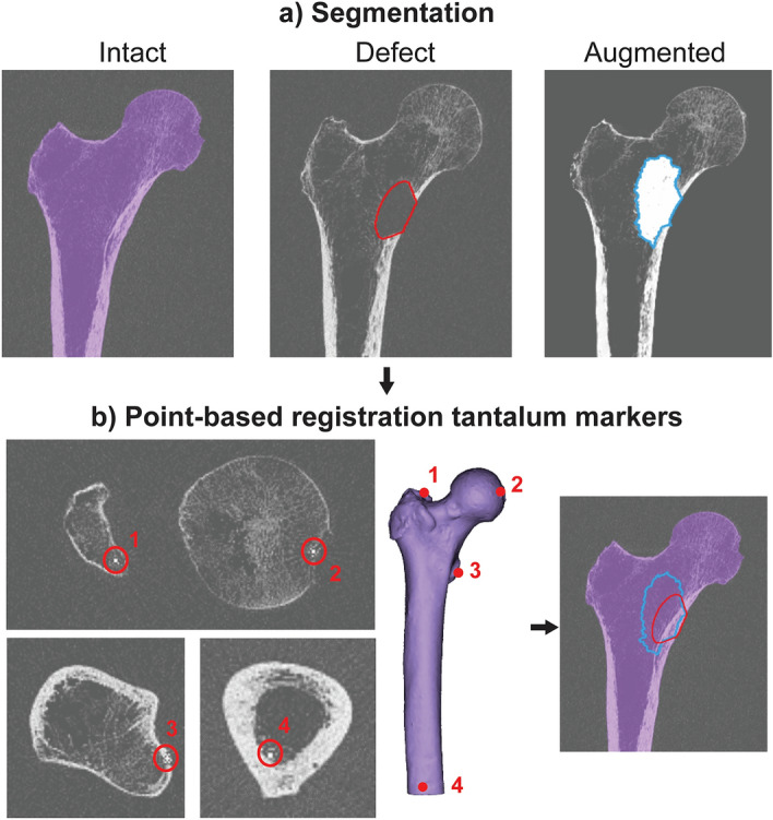
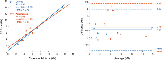
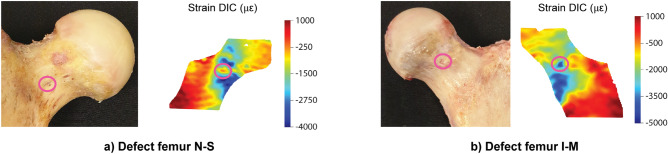
Femoroplasty is a procedure where bone cement is injected percutaneously into a weakened proximal femur. Uncertainty exists whether femoroplasty provides sufficient mechanical strengthening to prevent fractures in patients with femoral bone metastases. Finite element models are promising tools to evaluate the mechanical effectiveness of femoroplasty, but a thorough validation is required. This study validated a voxel-based finite element model against experimental data from eight pairs of human cadaver femurs with artificial metastatic lesions. One femur from each pair was left untreated, while the contralateral femur was augmented with bone cement. Finite element models accurately predicted the femoral strength in the defect (R2 = 0.96) and augmented (R2 = 0.93) femurs. The modelled surface strain distributions showed a good qualitative match with results from digital image correlation; yet, quantitatively, only moderate correlation coefficients were found for the defect (mean R2 = 0.78) and augmented (mean R2 = 0.76) femurs. This was attributed to the presence of vessel holes in the femurs and the jagged surface representation of our voxel-based models. Despite some inaccuracies in the surface measurements, the FE models accurately predicted the global bone strength and qualitative deformation behavior, both before and after femoroplasty. Hence, they can offer a useful biomechanical tool to assist clinicians in assessing the need for prophylactic augmentation in patients with metastatic bone disease, as well as in identifying suitable patients for femoroplasty.


Conflict of interest statement

The authors declare no competing interests.

Figures

Figure 1
Figure 1
Overview of the validation workflow. (a) Cadaver femurs were mechanically tested in a single leg stance configuration and displacements and strains were measured using digital image correlation (DIC). (b) CT scans of the femurs were resliced along the experimental coordinate system (blue) and downscaled to a 2 mm voxel size to enable direct conversion into a voxel-based finite element (FE) model mimicking the experimental conditions. (c) The FE models were validated by comparing the strength against the experimental failure load and the surface displacements and strains against the registered DIC measurements.
Figure 2
Figure 2
Segmentation and registration. (a) The femur, lesion and cement contour were segmented from the intact, defect and augmented CT scan respectively. (b) The lesion and cement contour were superimposed on the intact CT scan after a point-based registration of the four tantalum markers in each femur [at the top of the greater trochanter (1), the fovea of the femoral head (2), the lesser trochanter (3) and the posterior endosteal surface at the distal end of the femoral shaft (4)].
Figure 3
Figure 3
Validation failure force. A strong linear relation was found between the failure force quantified by FE and the experimental failure force, both for the defect and augmented group. Correlation graphs (left) and Bland–Altmann plots (right) are depicted.
Figure 4
Figure 4
Validation displacements. (Left) Qualitative comparison of the DIC and FE displacements at the anterior surface of the proximal femur. The results are displayed for one representative specimen pair (N–M) at 75% peak experimental force. Note that the scales for the FE and DIC measurements were not matched to allow a better visual comparison of the relative distribution. (Right) Quantitative comparison of the displacement values through linear regression analysis.
Figure 5
Figure 5
Validation strains. (Left) Qualitative comparison of the nominal DIC and FE strains at the anterior surface of the proximal femur. The results are displayed for a representative specimen pair (N–M) at 75% peak experimental force. Note that the scales for the FE and DIC measurements were not matched to allow a better visual comparison of the relative distribution. (Right) Quantitative comparison of the strain values through linear regression analysis.
Figure 6
Figure 6
Local strain concentrations at vessel holes. Two examples [defect femurs from pair N–S (a) and pair I–M (b)] illustrate how vessel holes can result in local strain concentrations in the DIC measurements.

Similar articles

Cited by

References

    1. Gendi K, Hennessy D, Heiner J. The burden of metastatic disease of the femur on the medicare system. Springerplus. 2016;5:1–10. doi: 10.1186/s40064-016-3572-8. - DOI - PMC - PubMed
    1. Mavrogenis AF, et al. Survival analysis of patients with femoral metastases. J. Surg. Oncol. 2012;105:135–141. doi: 10.1002/jso.22061. - DOI - PubMed
    1. Ratasvuori M, et al. Insight opinion to surgically treated metastatic bone disease: Scandinavian Sarcoma Group Skeletal Metastasis Registry report of 1195 operated skeletal metastasis. Surg. Oncol. 2013;22:132–138. doi: 10.1016/j.suronc.2013.02.008. - DOI - PubMed
    1. Philipp TC, Mikula JD, Doung YC, Gundle KR. Is there an association between prophylactic femur stabilization and survival in patients with metastatic bone disease? Clin. Orthop. Relat. Res. 2020;478:540–546. doi: 10.1097/CORR.0000000000000803. - DOI - PMC - PubMed
    1. Feng H, et al. CT-guided percutaneous femoroplasty (PFP) for the treatment of proximal femoral metastases. Pain Physician. 2016;19:E767–E773. - PubMed

Publication types