. 2022 May 9;15(1):43.
doi: 10.1186/s13041-022-00930-x.

CaVβ-subunit dependence of forward and reverse trafficking of CaV1.2 calcium channels

Affiliations

CaVβ-subunit dependence of forward and reverse trafficking of CaV1.2 calcium channels

Laurent Ferron et al. Mol Brain. .

Abstract

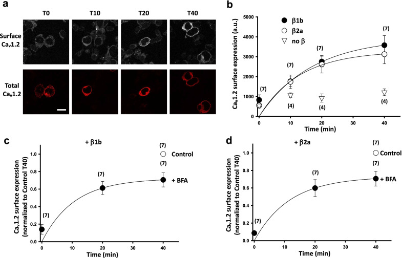
Auxiliary CaVβ subunits interact with the pore forming CaVα1 subunit to promote the plasma membrane expression of high voltage-activated calcium channels and to modulate the biophysical properties of Ca2+ currents. However, the effect of CaVβ subunits on channel trafficking to and from the plasma membrane is still controversial. Here, we have investigated the impact of CaVβ1b and CaVβ2a subunits on plasma membrane trafficking of CaV1.2 using a live-labeling strategy. We show that the CaVβ1b subunit is more potent in increasing CaV1.2 expression at the plasma membrane than the CaVβ2a subunit and that this effect is not related to modification of intracellular trafficking of the channel (i.e. neither forward trafficking, nor recycling, nor endocytosis). We conclude that the differential effect of CaVβ subunit subtypes on CaV1.2 surface expression is likely due to their differential ability to protect CaV1.2 from degradation.

Keywords: CaVβ auxiliary subunits; Calcium channel; Trafficking.


Conflict of interest statement

The authors declare no competing interest.

Figures

Fig. 1
Fig. 1
Effect of CaVβ subunits on CaV1.2 cell surface expression in tsA-201 cells. a Schematic of CaV1.2 channel tagged with bungarotoxin binding site (CaV1.2-BBS, BBS: red triangle) between S5 and the P-loop of domain II (DII). b Representative whole-cell current traces recorded in response to depolarizing steps from − 50 to + 40 mV from a holding potential of − 100 mV from tsA-201 cells expressing either CaV1.2 WT (top traces) or CaV1.2-BBS (bottom traces) together with auxiliary subunits CaVα2δ-1 and CaVβ1b. Mean I/V curves (right panel) for CaV1.2 WT (filled circle, n = 9) and CaV1.2-BBS (open circle, n = 18) co-expressed with auxiliary subunits CaVα2δ-1 and CaVβ1b. c Confocal images showing plasma membrane expression of CaV1.2-BBS in tsA cells stained with α-bungarotoxin (BTX)-AF488 (top panels). CaV1.2-BBS was co-expressed with CaVα2δ-1 (left, no β) and either CaVβ1b (center) or CaVβ2a (right). Cells were incubated at 17 °C with BTX-AF488 for 30 min and fixed. The cells were then permeabilized and stained with a rabbit anti-CaV1.2 Ab and secondary Ab anti-rabbit AF594 (bottom panels). Scale 20 µm. d Average CaV1.2-BBS surface expression co-transfected with CaVα2δ-1 and either CaVβ1b (black bar), or CaVβ2a (open bar), or empty vector (gray bar). Bars are mean (± SEM) normalized to CaVβ1b mean. ***p < 0.001, n = 14; $$$p < 0.001, n = 5; paired t-test, n numbers correspond to independent experiments
Fig. 2
Fig. 2
CaV1.2 endocytosis is dynamin-dependent and CaVβ subtype-independent. a Representative confocal images of tsA-201 cells expressing CaV1.2-BBS and labelled with BTX-AF488 (top panels). CaV1.2-BBS was co-expressed with CaVα2δ-1 and CaVβ1b. Cells were incubated at 17 °C with BTX-AF488 for 30 min and then fixed at different time point after incubation at 37 °C, from zero (T0) to 20 min (T20). The cells were then permeabilized and stained with a rabbit anti-CaV1.2 Ab and a secondary Ab anti-rabbit AF594 (bottom panels). Scale bar 20 µm. b Time course of endocytosis of cell surface CaV1.2-BBS co-expressed with CaVα2δ-1 and either CaVβ1b (filled circle) or CaVβ2a (open circle). The results are shown as the mean ± SEM. The n numbers correspond to independent experiments (average fluorescence from at least 25 cells per time point). The data were fitted with single exponentials. The time constants of the fits were 5.6 ± 0.2 min for CaVβ1b and 7.1 ± 0.2 min for CaVβ2a, respectively. c and d Effect of dominant negative dynamin Dyn K44E on CaV1.2 endocytosis. Cells were transfected with CaV1.2-BBS, CaVα2δ-1 and either CaVβ1b (c) or CaVβ2a (d) together with either empty pcDNA3.1 vector (filled symbols) or Dyn K44E (DDN, open symbols). Cells were incubated at 17 °C with BTX-AF488 for 30 min and then fixed at time point T0 and T20 after incubation at 37 °C. Cells were subjected to immunocytochemistry as described in a. BTX-AF488 fluorescence was normalized to the mean fluorescence at T0 for each condition. The results are shown as the mean ± SEM. The n numbers correspond to independent experiments (average fluorescence from at least 25 cells per time point). $$p < 0.01 Control T10 vs Control T0; *p < 0.05 DDN T10 vs Control T10, unpaired t-test
Fig. 3
Fig. 3
CaV1.2 forward trafficking and recycling are not CaVβ-subtype dependent. a Confocal images of tsA-201 cells expressing CaV1.2-BBS and labelled with BTX-AF488 (top panels). CaV1.2-BBS was co-expressed with CaVα2δ-1 and CaVβ1b. Cells were incubated at 17 °C with untagged BTX for 30 min and then incubated at 37 °C with BTX-AF488. The cells were fixed at different time point after incubation at 37 °C, from zero (T0) to 40 min (T40). The cells were then permeabilized and stained with a rabbit anti-CaV1.2 Ab and a secondary Ab anti-rabbit AF594 (bottom panels). Scale bar 20 µm. b Time course of insertion of CaV1.2-BBS at the cell surface when co-expressed with CaVα2δ-1 and either CaVβ1b (filled circle), CaVβ2a (open circle) or empty vector (open triangle). The results are shown as the mean ± SEM (n numbers correspond to independent experiments). Data were fitted with single exponentials. The time constants of the fits were 16.8 ± 12.6 min and 13.0 ± 14.2 min for CaVβ1b and CaVβ2a (n = 7), respectively. c and d Effect of Brefeldin A (BFA) treatment on CaV1.2 forward trafficking. tsA-201 cells were co-transfected with CaV1.2-BBS, CaVα2δ-1 and either CaVβ1b (c) or CaVβ2a (d). Cells were treated with BFA for 4 h before undergoing the forward trafficking protocol described in a. BTX-AF488 fluorescence was normalized to the mean fluorescence at T40 for the control condition (open circle). The results are shown as the mean ± SEM. The n numbers correspond to independent experiments (average fluorescence from at least 25 cells per time point). The data were compared using an unpaired t-test. The data were fitted with single exponentials. The time constants of the fits were 10.6 ± 6.6 min and 11.6 ± 8.1 min for CaVβ1b and CaVβ2a (n = 7), respectively

Similar articles

Cited by

References

    1. Catterall WA, Leal K, Nanou E. Calcium channels and short-term synaptic plasticity. J Biol Chem. 2013;288:10742–10749. doi: 10.1074/jbc.R112.411645. - DOI - PMC - PubMed
    1. Wheeler DG, et al. Ca(V)1 and Ca(V)2 channels engage distinct modes of Ca(2+) signaling to control CREB-dependent gene expression. Cell. 2012;149:1112–1124. doi: 10.1016/j.cell.2012.03.041. - DOI - PMC - PubMed
    1. Catterall WA, Lenaeus MJ, Gamal El-Din TM. Structure and pharmacology of voltage-gated sodium and calcium channels. Annu Rev Pharmacol Toxicol. 2020;60:133–154. doi: 10.1146/annurev-pharmtox-010818-021757. - DOI - PubMed
    1. Zamponi GW, Striessnig J, Koschak A, Dolphin AC. The physiology, pathology, and pharmacology of voltage-gated calcium channels and their future therapeutic potential. Pharmacol Rev. 2015;67:821–870. doi: 10.1124/pr.114.009654. - DOI - PMC - PubMed
    1. Obermair GJ, Szabo Z, Bourinet E, Flucher BE. Differential targeting of the L-type Ca2+ channel alpha 1C (CaV1.2) to synaptic and extrasynaptic compartments in hippocampal neurons. Eur J Neurosci. 2004;19:2109–2122. doi: 10.1111/j.0953-816X.2004.03272.x. - DOI - PubMed

Publication types

MeSH terms

LinkOut - more resources