

Contrast-enhanced weighted-T1 and FLAIR sequences in MRI of meningeal lesions
- PMID: 35535121
- PMCID: PMC9077169
Contrast-enhanced weighted-T1 and FLAIR sequences in MRI of meningeal lesions
Abstract
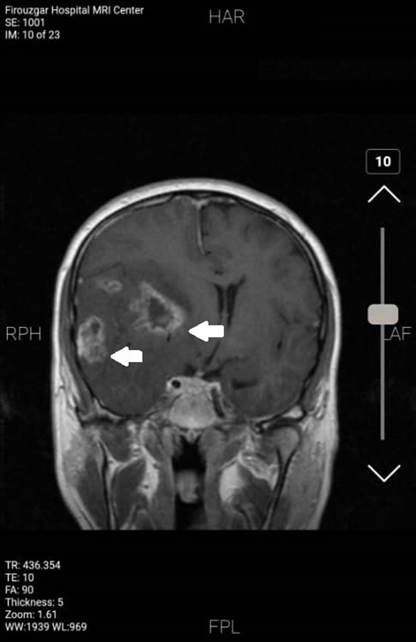
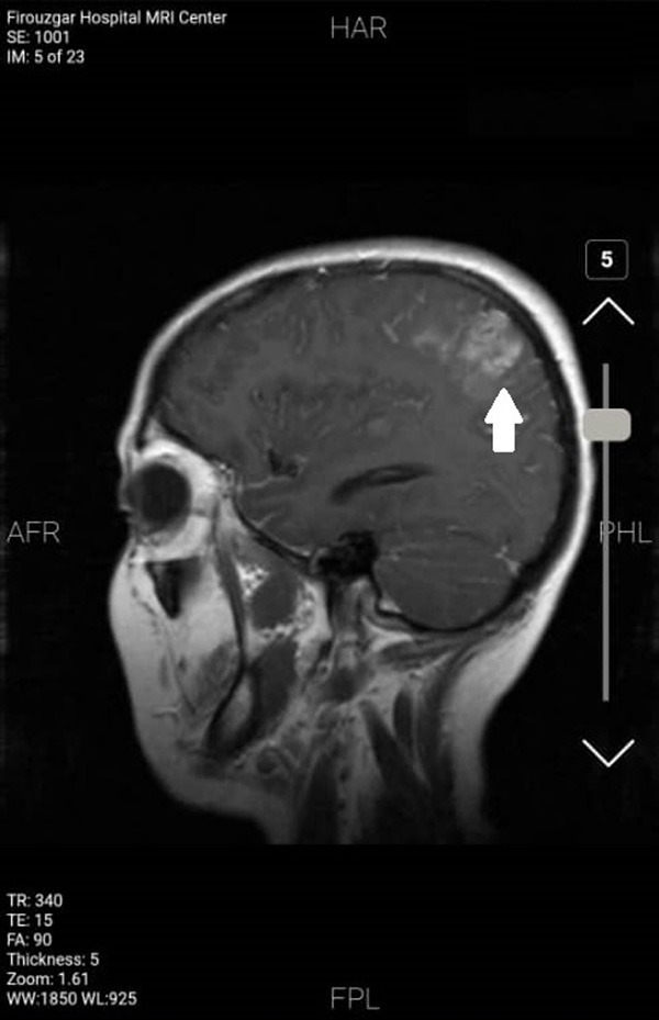
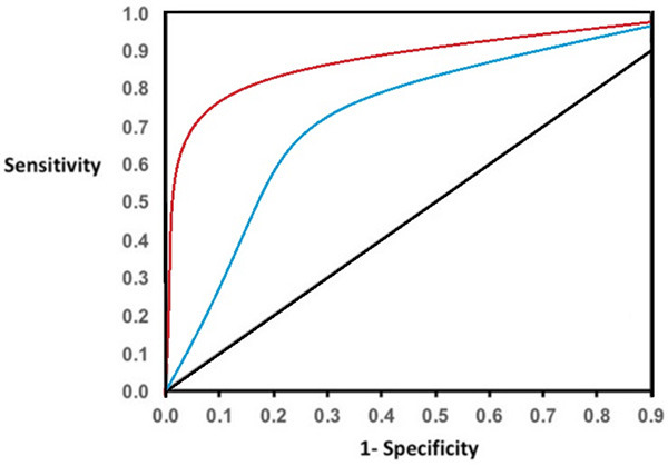
Magnetic resonance imaging (MRI) is widely used in meningeal lesions due to rapid and accurate diagnosis and prevention of serious complications. The aim of the present study was to compare these two sequences after injection of a contrast agent into meningeal lesions. This is a descriptive-analytical study that was performed in 2018-2020 on patients referred to the radiology ward with detection of any meningeal involvements in the MRI images. In addition to T1-W, FLAIR sequence imaging was also performed. Images were initially evaluated by two expert radiologists and a neurologist. The diagnostic values of the sequences were compared. Overall, a total number of 147 patients with meningeal lesions in their brain MRI entered the study. 57.1% of cases (84 patients) had an infectious etiology and 42.9% (63 patients) had a tumoral etiology. T1-W images without contrast were able to diagnose 78 cases of meningitis (92.8% of them), and FLAIR sequences could diagnose 82 patients (97.6% of them). Without contrast injection on MRI, the diagnostic value of T1-W sequence was higher than FLAIR sequence for tumoral lesions (P < 0.01). The enhancement degree of T1-W was higher for tumoral findings (P < 0.01). In contrast, the enhancement degree of the FLAIR sequence was higher for infectious findings, which was also statistically significant (P = 0.015). FLAIR sequences had 92% sensitivity and 85% specificity for diagnosis of brain inflammatory diseases. Similar analysis showed that T1 sequence had 82% sensitivity and 73% specificity for diagnosis of brain inflammatory diseases.
Keywords: FLAIR; MRI; T1-W; meningeal lesions.
AJNMMI Copyright © 2022.
Conflict of interest statement
None.
Figures
References
-
- Choi SJ, Park YH, Kim JA, Han JH, Choe G, Kim S. Pearls & oy-sters: asymmetric meningeal involvement is a common feature of rheumatoid meningitis. Neurology. 2017;88:e108–e110. - PubMed
-
- Magill ST, Young JS, Chae R, Aghi MK, Theodosopoulos PV, McDermott MW. Relationship between tumor location, size, and WHO grade in meningioma. Neurosurg Focus. 2018;44:E4. - PubMed
-
- Apra C, Peyre M, Kalamarides M. Current treatment options for meningioma. Expert Rev Neurother. 2018;18:241–249. - PubMed
LinkOut - more resources
Full Text Sources