. 2022 Jan 29:17:100431.
doi: 10.1016/j.ynstr.2022.100431. eCollection 2022 Mar.

Sex and chronic stress alter the distribution of glutamate receptors within rat hippocampal CA3 pyramidal cells following oxycodone conditioned place preference

Affiliations

Sex and chronic stress alter the distribution of glutamate receptors within rat hippocampal CA3 pyramidal cells following oxycodone conditioned place preference

Alexandra Dolgetta et al. Neurobiol Stress. .

Abstract

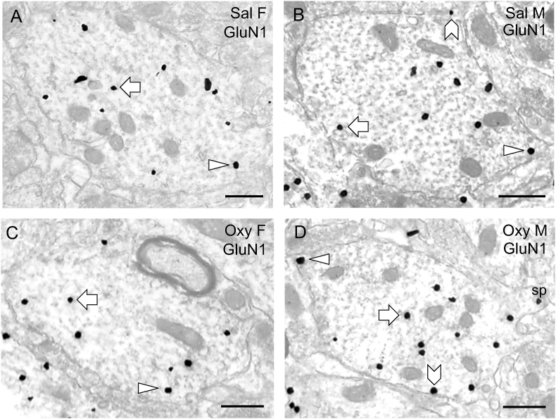
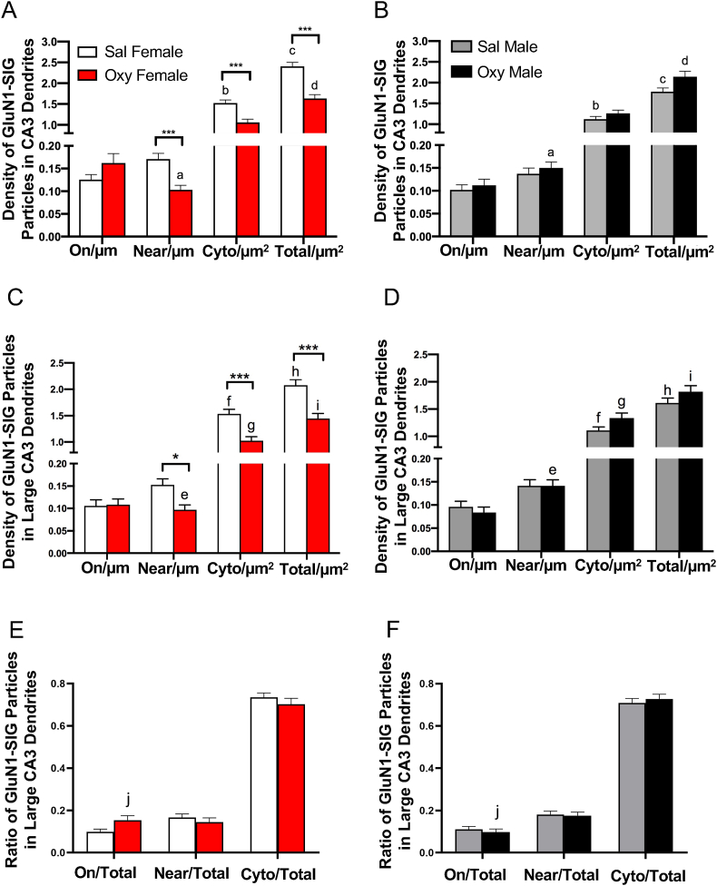
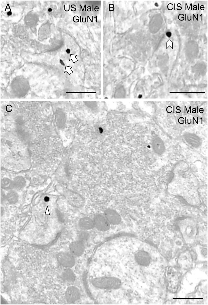
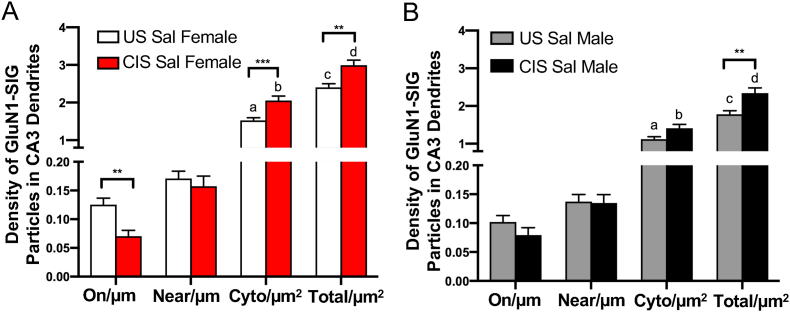
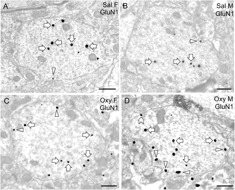
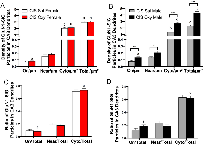
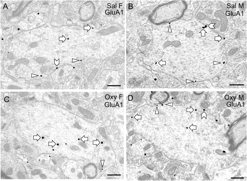
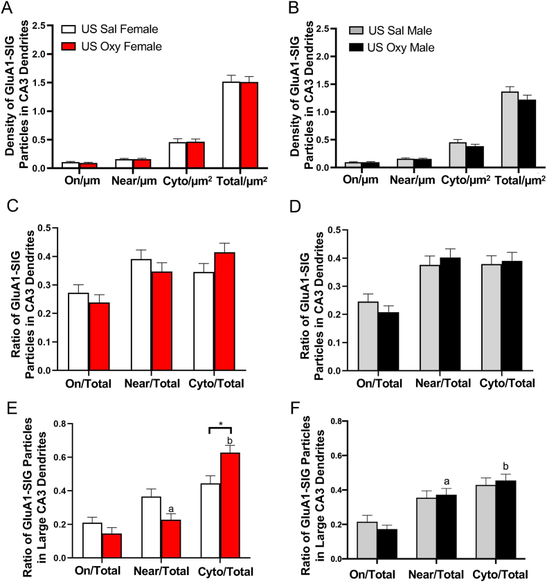
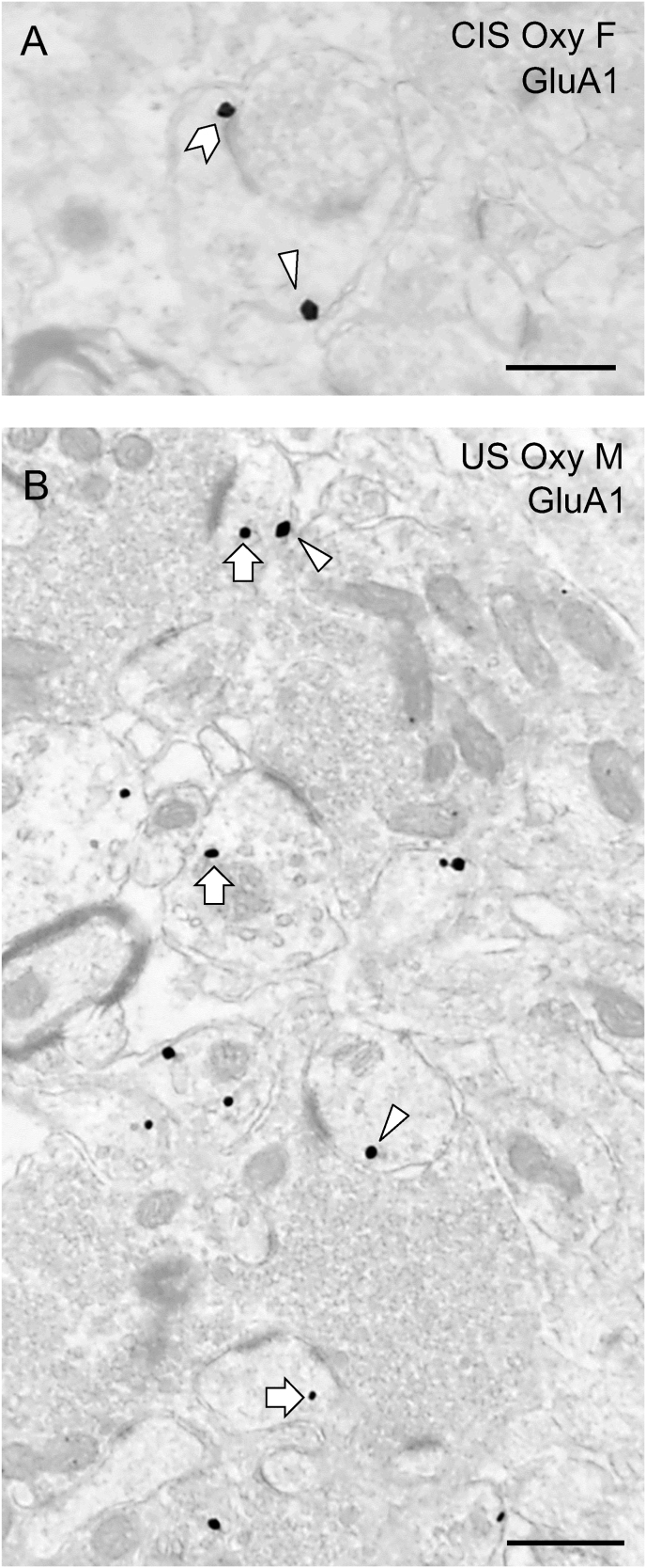
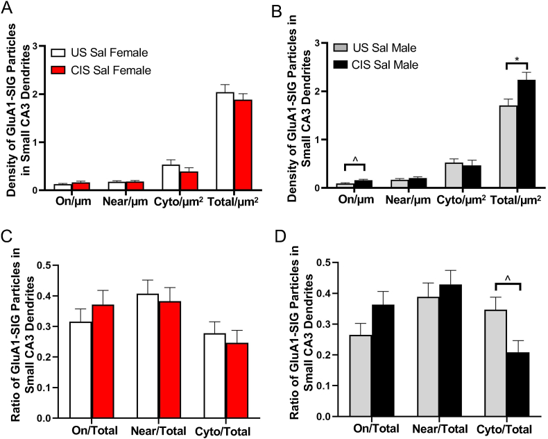
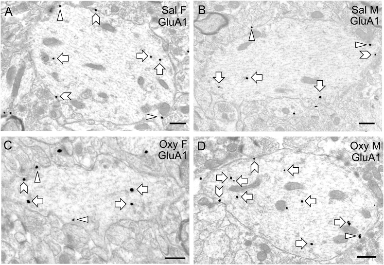
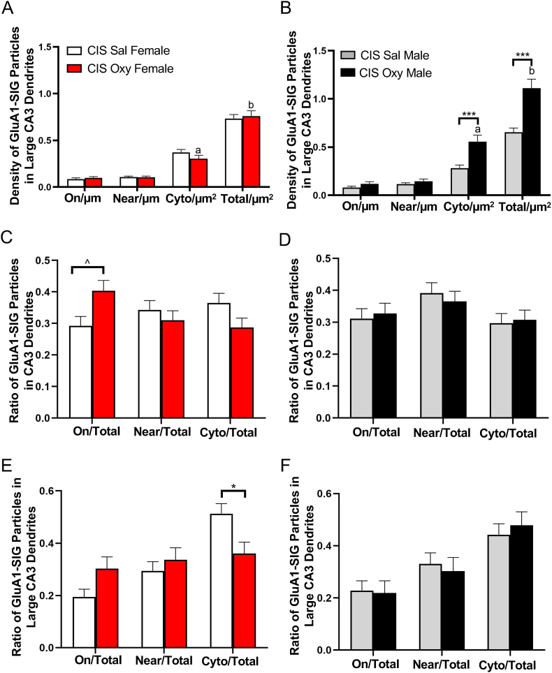
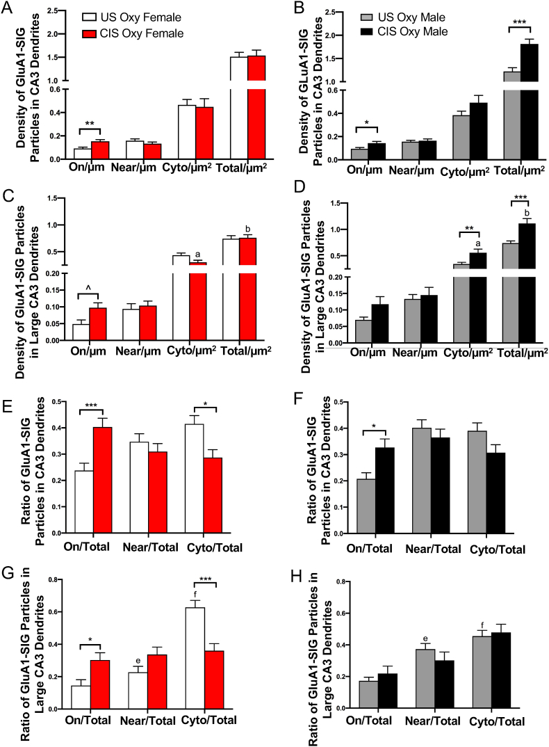
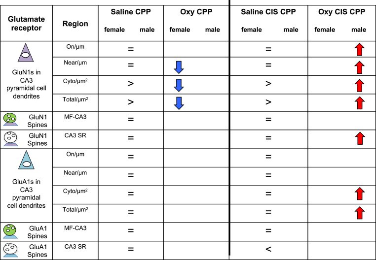
Glutamate receptors have a key role in the neurobiology of opioid addiction. Using electron microscopic immunocytochemical methods, this project elucidates how sex and chronic immobilization stress (CIS) impact the redistribution of GluN1 and GluA1 within rat hippocampal CA3 pyramidal cells following oxycodone (Oxy) conditioned place preference (CPP). Four groups of female and male Sprague-Dawley rats subjected to CPP were used: Saline- (Sal) and Oxy-injected (3 mg/kg, I.P.) naïve rats; and Sal- and Oxy-injected CIS rats. GluN1: In both naive and CIS rats, Sal-females compared to Sal-males had elevated cytoplasmic and total dendritic GluN1. Following Oxy CPP, near plasmalemmal, cytoplasmic, and total GluN1 decreased in CA3 dendrites of unstressed females suggesting reduced pools of GluN1 available for ligand binding. Following CIS, Oxy-males (which did not acquire CPP) had increased GluN1 in all compartments of dendrites and spines of CA3 neurons. GluA1: There were no differences in the distribution GluA1 in any cellular compartments of CA3 dendrites in naïve females and males following either Sal or Oxy CPP. CIS alone increased the percent of GluA1 in CA3 dendritic spines in males compared to females. CIS Oxy-males compared to CIS Sal-males had an increase in cytoplasmic and total dendritic GluA1. Thus, in CIS Oxy-males increased pools of GluN1 and GluA1 are available for ligand binding in CA3 neurons. Together with our prior experiments, these changes in GluN1 and GluA1 following CIS in males may contribute to an increased sensitivity of CA3 neurons to glutamate excitation and a reduced capacity to acquire Oxy CPP.

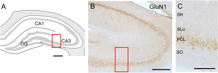
Keywords: ABC, avidin-biotin complex; AMPA receptors; BSA, bovine serum albumin; CIS, chronic immobilization stress; CPP, conditioned place preference; DAB, diaminobenzidine; DG, dentate gyrus; DOR, delta opioid receptor; Drug associative-learning; Electron microscopy; GABA, Gamma-amino butyric acid; GluA1, AMPA glutamate receptor subunit 1; GluN1, NMDA, glutamate receptor subunit 1; LTP, long-term potentiation; MOR, mu opioid receptor; NMDA receptors; NMDA, N-methyl-D-aspartate; NPY, neuropeptide Y; Oxy, oxycodone; PARV, parvalbumin; PB, phosphate buffer; PFA, paraformaldehyde; PM, plasma membrane; Pyramidal cells; ROI, region of interest; SLM, stratum lacunosum-moleculare; SLu, stratum lucidum; SO, stratum oriens; SOM, somatostatin; SR, stratum radiatum; Sal, saline; TS, tris-buffered saline; ir, immunoreactivity.


Conflict of interest statement

The authors declare no competing financial interests.

Figures

Fig. 1
Fig. 1
Hippocampal region sampled for electron microscopy. A. Schematic diagram of the rostral rat hippocampus showing the CA1, CA3 and dentate gyrus subregions. CA3 (boxed region) was sampled for electron microscopy [modified from diagram 31 (−3.70 from bregma) in (Swanson, 1992)]. B. By light microscopy, dense GluN1-immunoreactivity is seen in the pyramidal cell layer (PCL) and to a lesser extent in stratum oriens (SO), stratum lucidum (SLu) and stratum radiatum (SR). Scale bar = 0.5 mm.
Fig. 2
Fig. 2
Representative electron micrographs of GluN1-SIG particles in CA3 pyramidal cell dendrites in naïve Sal- and Oxy-female and male rats. A-D. Electron micrographs show the distribution of GluN1-SIG particles within dendrites from a Sal-female (A), a Sal-male (B), an Oxy-female (C), and an Oxy-male (D) rat. Examples of near plasmalemmal (triangle) and cytoplasmic (arrow) DOR-SIG particles in dendrites are shown. Scale bar = 500 nm.
Fig. 3
Fig. 3
Quantitative analysis of the distribution of GluN1-SIG particles in CA3 pyramidal cell dendrites in naïve Sal- and Oxy-female and male rats. A, B. Sal-females compared to Sal-males have a greater density of GluN1-SIG particles in the cytoplasm and in total in CA1 pyramidal cell dendrites. Sal-females compared to Oxy-females had greater GluN1-SIG particles near the plasma membrane, in the cytoplasm, and in total in CA3 dendrites (A). Oxy-females compared to Oxy-males have a decreased density of GluN1-SIG particles near the plasmalemma and in total. C, D. When dendrites are divided on the basis of size, Sal-females compared to Sal-males have a greater density of GluN1-SIG particles in the cytoplasm and in total in large CA3 dendrites. Sal-females compared to Oxy-females have a greater density of GluN1-SIG particles near the plasma membrane, in the cytoplasm, and in total in large CA3 dendrites (C). Oxy-females compared to Oxy-males have a decreased density of GluN1-SIG particles near the plasma membrane, in the cytoplasm, and in total in large CA3 dendrites. E, F. Oxy-females compared to Oxy-males have a greater ratio of GluN1-SIG particles in large CA3 dendrites. ***p < 0.001,**p < 0.01, *p < 0.05, pb,c,f < 0.001, pd,h < 0.01, pa,g,i < 0.05, pe = 0.05, N = 3 rats per group; n = 50 dendrites per rat.
Fig. 4
Fig. 4
Examples of GluN1-labeled spines in CA3. Usually one GluN1-SIG particle was found in a single dendritic CA3 spine, but sometimes more than one was detected. A. A CA3 spine in the SR of an unstressed (US) male contains two GluN1-SIG particles in the cytoplasm (arrows). B. A GluN1-SIG particle on the synapse (chevron) in a SR spine of a CIS male. Both spines are contacted by unlabeled terminals (uT) C. A GluN1-SIG particle is found near the plasma membrane (arrowhead) of a CA3 spine contacted by a mossy fiber (MF) in the SLu. Scale bar: 500 nm.
Fig. 5
Fig. 5
Quantitative analysis of the distribution of GluN1-SIG particles in CA3 pyramidal cell dendrites in unstressed (US) and CIS Sal-female and Sal-male rats. A,B. US Sal-females compared to US Sal-males have a greater density of GluN1-SIG particles in the cytoplasm and in total in CA3 dendrites. After undergoing CIS, females (A) have a greater density of GluN1-SIG particles in the cytoplasm and in total in CA3 dendrites, but a decreased density of GluN1-SIG particles on the plasma membrane. After undergoing CIS, males (B) have a greater density of GluN1-SIG particles in total in CA3 dendrites. CIS Sal-females compared to CIS Sal-males have a greater density of GluN1-SIG particles in the cytoplasm and in total in CA3 dendrites. ***p < 0.001,**p < 0.01, *p < 0.05, Pa < 0.05, pb,c,d < 0.001. N = 3 rats per group; n = 50 dendrites per rat.
Fig. 6
Fig. 6
Representative electron micrographs showing GluN1-SIG labeling in CA3 pyramidal cell dendrites for CIS Sal- and Oxy-female and male rats. A-D. Electron micrographs show the distribution of GluN1-SIG particles within CA3 pyramidal cell dendrites from CIS Sal-female (A), a CIS Sal-male (B), a CIS Oxy-female (C), and a CIS Oxy-male (D) rat. Examples of near plasmalemmal (triangle), on plasmalemmal (chevron), and cytoplasmic (arrow) GluN1-SIG particles in dendrites are shown. Scale bar: 500 nm.
Fig. 7
Fig. 7
Quantitative analysis of the distribution of GluN1-SIG particles in CA3 pyramidal cell dendrites in CIS Sal- and Oxy-female and male rats. A, B. CIS Sal-females compared to CIS Sal-males have a greater density of GluN1-SIG particles in the cytoplasm and in total in CA3 dendrites. In CIS females, there is no effect of Oxy CPP on the density of GluN1-SIG particles (A). However, CIS Oxy-males compared to CIS Sal-males (B) have a greater density of GluN1-SIG particles on and near the plasmalemma, in the cytoplasm, and in total in CA3 dendrites. CIS Oxy-females compared to CIS Oxy-males have a lower density of GluN1-SIG particles on the plasmalemma, in the cytoplasm, and in total in CA3 dendrites. C, D. CIS Oxy-females compared to CIS Oxy-males had a significantly lower ratio of GluN1-SIG particles on the plasma membrane, but a higher ratio of GluN1-SIG particles in the cytoplasm. ***p < 0.001, **p < 0.01, *p < 0.05, pf < 0.001, pa,b < 0.01, pa,b,c,e,g < 0.05. N = 3 rats per group; n = 50 dendrites per rat.
Fig. 8
Fig. 8
Representative electron micrographs of GluA1-SIG particles in CA3 pyramidal cell dendrites in naïve Sal- and Oxy-female and male rats. A-D. Electron micrographs show the distribution of GluA1-SIG particles within dendrites from a Sal-female (A), a Sal-male (B), an Oxy-female (C), and an Oxy-male (D) rat. Examples of near plasmalemmal (triangle) and cytoplasmic (arrow) GluA1-SIG particles in dendrites are shown. Scale bar = 500 nm.
Fig. 9
Fig. 9
Quantitative analysis of the distribution of GluA1-SIG particles in CA3 pyramidal cell dendrites in naïve Sal- and Oxy-female and male rats. A-B There were no observed within or across sex differences in the distribution of GluA1-SIG particles in CA3 pyramidal cell dendrites. C, D. There were no observed within or across sex differences in the partitioning ratios of GluA1 in any cellular compartment of CA3 pyramidal cell dendrites. E, F. When specifically selecting for large dendrites, treatment and sex differences were revealed. US Oxy-females compared to US Sal-females had a significantly greater ratio of GluA1-SIG particles in the cytoplasm of large CA3 dendrites. Moreover, US Oxy-males compared to US Oxy-females have a greater ratio of GluA1-SIG particles near the cytoplasm, but a smaller ratio of GluA1-SIG particles in the cytoplasm., *p < 0.05, pa = 0.05, pb < 0.05. N = 3 rats per group; n = 50 dendrites per rat.
Fig. 10
Fig. 10
Examples of GluA1-labeled spines in CA3. Usually one GluA1-SIG particle was found in a single dendritic CA3 spine, but sometimes more than one was detected. A. A SR spine of a CIS Oxy-male has GluA1-SIG particles on the synapse (chevron) and near the plasma membrane (arrowhead). B. Numerous dendritic spines containing GluA1-SIG particles and contacted by mossy fibers are observed in SLu. Examples of near plasma membrane (arrowhead) and cytoplasmic (arrows) GluA1-SIG particles are shown. Scale bar: 500 nm.
Fig. 11
Fig. 11
Quantitative analysis of the distribution of GluA1-SIG particles in small CA3 pyramidal cell dendrites in unstressed (US) and CIS Sal-female and Sal-male rats. A-D. There are no sex differences between US or CIS Sal-female or Sal-male rats when comparing density or partitioning ratio of GluA1-SIG particles in small CA3 dendrites. Furthermore, there are no differences observed between US and CIS Sal-female rats for density (A) and portioning ratio (C) of GluA1-SIG particles in small CA3 dendrites. There are, however, significant differences between US Sal-male and CIS Sal-male rats in GluA1-SIG particles density (B) and partitioning ratio (D) in small CA3 dendrites. B. CIS males compared to US males had greater density of GluA1-SIG particles on the plasma membrane and in total in small CA3 dendrites. D. Moreover, the ratio of GluA1-SIG particles in the cytoplasm of small CA3 dendrites was greater in US males than CIS males. *p < 0.05, ^p = 0.06. N = 3 rats per group; n = 50 dendrites per rat.
Fig. 12
Fig. 12
Representative electron micrographs showing GluA1-SIG labeling in CA3 pyramidal cell dendrites for CIS Sal- and Oxy-female and male rats. A-D. Electron micrographs show the distribution of GluA1-SIG particles within CA3 pyramidal cell dendrites from a CIS Sal-female (A), a CIS Sal-male (B), a CIS Oxy-female (C), and a CIS Oxy-male (D) rat. Examples of near plasmalemmal (triangle), on plasmalemmal (chevron), and cytoplasmic (arrow) GluN1-SIG particles in dendrites are shown. Scale bar: 500 nm.
Fig. 13
Fig. 13
Quantitative analysis of the distribution of GluA1-SIG particles in CA3 pyramidal cell dendrites in CIS Sal- and Oxy-female and male rats. A, B. There were no sex differences between CIS Sal-female and Sal-male rats. However, there were significant differences between CIS Oxy-female and CIS Oxy-male rats, with CIS-male rats demonstrating greater density of GluA1-SIG particles in the cytoplasm and in total in large CA3 dendrites. Moreover, while there were no treatment differences between CIS Oxy- and Sal-female rats (A), there were differences observed between CIS Oxy- and Sal-male rats. CIS Oxy-male rates compared to CIS Sal-male rates had a greater density of GluA1-SIG particles in the cytoplasm and in total in large CA3 dendrites (B). C–F. There were no significant sex differences between CIS Sal- or Oxy-female or male rats in the partitioning ratio of GluA1-SIG particles in CA3 of all sizes or specifically in large CA3 dendrites. However, there were significant treatment differences specifically between CIS Sal- and Oxy-female, not male (D, F), rats in the partitioning ratio of GluA1-SIG particles in both CA3 dendrites of all sizes (C) and large CA3 dendrites (E). CIS Oxy-females as opposed to CIS Sal-females had a greater ratio of GluA1-SIG particles on the plasmalemma across CA3 dendrites of all sizes. When specifically examining large CA3 dendrites, CIS Oxy-females as compared to CIS Sal-females had a decreased ratio of GluA1-SIG particles in the cytoplasm. ***p < 0.001, *p < 0.05, ^p = 0.06, pa,b < 0.001. N = 3 rats per group; n = 50 dendrites per rat.
Fig. 14
Fig. 14
Quantitative analysis of the distribution of GluA1-SIG particles in CA3 pyramidal dendrites in unstressed (US) and CIS Oxy-female and Oxy-male rats. A-D. There were no observed sex differences between US Oxy-female and Oxy-male in the density of GluA1-SIG particles in CA3 dendrites of all sizes or specifically within large CA3 dendrites. After undergoing CIS Oxy-females had a greater density of GluA1-SIG particles on the plasmalemma in CA3 dendrites of all sizes and specifically in large CA3 dendrites (A, C). After undergoing CIS Oxy-males had a greater density of GluA1-SIG particles on the plasmalemma and in total in CA3 dendrites of all sizes, and a greater density of GluA1-SIG particles on the plasmalemma, in the cytoplasm, and in total in large CA3 dendrites (B, D). When examining partitioning ratios, CIS Oxy-females compared to US Oxy-females had a greater ratio of GluA1-SIG particles on the plasmalemma in large CA3 dendrites and CA3 dendrites of all sizes, but a decreased ratio of GluA1-SIG particles in the cytoplasm of large CA3 dendrites and CA3 dendrites of all sizes. CIS Oxy-males as compared to US Oxy-males had an increased ratio of GluA1-SIG particles on the plasmalemma of CA3 dendrites of all sizes (F), but no observed differences in partitioning ratios in large CA3 dendrites (H). A-B. CIS Oxy-males as compared to CIS Oxy-females had a greater density of GluA1-SIG particles in the cytoplasm and in total in CA3 dendrites of all sizes and in large CA3 dendrites. However, US Oxy-female rats compared to US Oxy-male rats have a decreased ratio of GluA1-SIG particles near the plasmalemma and a greater ratio of GluA1-SIG particles in the cytoplasm specifically in large CA3 dendrites (GH), an effect which is not observed when examining CA3 dendrites of all sizes (EF). ***p < 0.001,**p < 0.01, *p < 0.05, ^p < 0.06, pb < 0.001, pa < 0.01, pc = 0.001, pd = 0.008, pf < 0.05, pe = 0.06. N = 3 rats per group; n = 50 dendrites per rat.
Fig. 15
Fig. 15
Summary diagram: sex differences in glutamate receptor subunit distribution in the hippocampal CA3 region. At baseline, Sal-females compared to Sal-males had greater densities of cytoplasmic and total GluN1 in CA3 pyramidal cell dendrites. Similarly, CIS Sal-females compared to CIS Sal-males had greater densities of cytoplasmic and total GluN1 in CA3 pyramidal cell dendrites. In contrast, there were no baseline differences in GluA1 in Sal-females compared to Sal-males in CA3 pyramidal cell dendrites or spines. Moreover, CIS Sal-females compared to CIS Sal-males had no baseline differences in GluA1 in CA3 pyramidal cell dendrites. However, unlike GluN1, GluA1 was lower in spines in CIS Sal-females compared to CIS Sal-males. Following Oxy CPP, GluN1 decreased near the plasma membrane, in the cytoplasm and in total in CA3 pyramidal cell dendrites in females only. In contrast, CIS Oxy-males (which did not acquire CPP) had elevated GluN1 and GluA1 in several compartments in CA3 pyramidal cell dendrites. CIS Oxy-males also had elevated numbers of GluN1-labeled spines in SR.

Similar articles

Cited by

  • Special issue dedicated to Dr. Bruce S. McEwen.
    Hill M, Hunter R, Reagan L. Hill M, et al. Neurobiol Stress. 2023 Jun 6;25:100552. doi: 10.1016/j.ynstr.2023.100552. eCollection 2023 Jul. Neurobiol Stress. 2023. PMID: 37547772 Free PMC article. No abstract available.

References

    1. Akashi K., Kakizaki T., Kamiya H., Fukaya M., Yamasaki M., Abe M., Natsume R., Watanabe M., Sakimura K. NMDA receptor GluN2B (GluR epsilon 2/NR2B) subunit is crucial for channel function, postsynaptic macromolecular organization, and actin cytoskeleton at hippocampal CA3 synapses. J. Neurosci. 2009;29:10869–10882. - PMC - PubMed
    1. Akil H., Gordon J., Hen R., Javitch J., Mayberg H., McEwen B., Meaney M.J., Nestler E.J. Treatment resistant depression: a multi-scale, systems biology approach. Neurosci. Biobehav. Rev. 2018;84:272–288. - PMC - PubMed
    1. American Addiction Centers . American Addiction Centers; 2020. How Hormones Affect Addiction.
    1. Ashirova E., Contoreggi N.H., Johnson M.A., Al-Khayat F.J., Calcano G.A., Rubin B.R., O'Cinneide E.M., Zhang Y., Zhou Y., Gregoire L., McEwen B.S., Kreek M.J., Milner T.A. Oxycodone injections not paired with conditioned place preference have little effect on the hippocampal opioid system in female and male rats. Synapse. 2021;75 - PubMed
    1. Barkóczi B., Juhász G., Averkin R.G., Vörös I., Vertes P., Penke B., Szegedi V. GluA1 phosphorylation alters evoked firing pattern in vivo. Neural Plast 2012. 2012:286215. - PMC - PubMed