

Case Reports
doi: 10.1155/2022/2953579.
eCollection 2022.
Massive Facial Presentation of Dermatofibrosarcoma Protuberans
Affiliations
- PMID: 35535304
- PMCID: PMC9078855
- DOI: 10.1155/2022/2953579
Item in Clipboard
Case Reports
Massive Facial Presentation of Dermatofibrosarcoma Protuberans
Case Rep Radiol.
.
Abstract
Dermatofibrosarcoma protuberans is a low-grade cutaneous sarcoma typically located on the trunk or proximal extremities. Less common locations include the head, face, and neck area. This tumour is slow growing with variable clinical appearance. It is known for its locally invasive nature and low metastatic propensity. Because imaging findings are rather nonspecific, biopsy is needed for definite diagnosis. This case describes an unusually large example of dermatofibrosarcoma protuberans in the less common preauricular region.
Copyright © 2022 Sebastiaan Hermans et al.
Conflict of interest statement
All authors declare no conflict of interest.
Figures
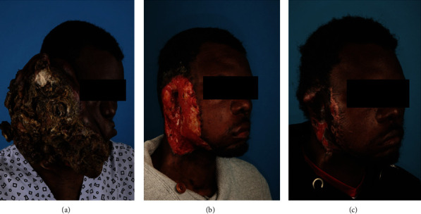
Photographs of a large right-sided facial necrotizing mass causing facial disfigurement after successive treatment. (a) Initial presentation. (b) After reconstruction of first tumour debulking with cutaneous allograft. (c) After reconstruction of the second debulking with split-thickness skin graft.
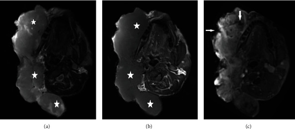
Axial MRI of the head showing a large lobulated mass centered at the right preauricular region. (a) The lesion appearing hypointense on T1-weighted imaging, (b) moderately hyperintense on T2-weighted imaging, and (c) showing marked homogeneous contrast enhancement with peripheral and intratumoral zones of necrosis (white arrows) on T1-weighted imaging after gadolinium administration. The masseter muscle and perioral muscles are not separately visible due to deep muscular invasion. There is also deep invasion of the tumour behind the right mandibular ramus in the masticator space.
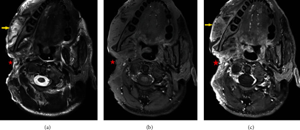
Axial MRI of the head showing postoperative outcome by after the first stage of debulking with a drastic reduction in tumour tissue (red star). (a) T2-weighted imaging, (b) T1-weighted imaging, and (c) T1-weighted imaging after gadolinium injection. The masticator muscles show increased T2w intensity and diffuse enhancement, due to muscle oedema (yellow arrow). Residual tumour tissue is difficult to differentiate for which follow-up imaging is necessary.
References
-
- Kumar L., Vimal Bhandari V., Singh S., Garg P., Kumar A. A giant dermatofibrosarcoma protuberans: a rare presentation over face. Journal of Cancer Research and Therapeutics . 2016;11(4):p. 1038. - PubMed
Publication types
LinkOut - more resources
Full Text Sources
