Perioperative Management and Long-Term Outcomes in Ocular Cicatricial Pemphigoid Patients Undergoing Cataract Surgery
- PMID: 35535356
- PMCID: PMC9078804
- DOI: 10.1155/2022/2496649
Perioperative Management and Long-Term Outcomes in Ocular Cicatricial Pemphigoid Patients Undergoing Cataract Surgery
Retraction in
-
Retracted: Perioperative Management and Long-Term Outcomes in Ocular Cicatricial Pemphigoid Patients Undergoing Cataract Surgery.Oxid Med Cell Longev. 2023 Sep 27;2023:9826796. doi: 10.1155/2023/9826796. eCollection 2023. Oxid Med Cell Longev. 2023. PMID: 37810560 Free PMC article.
Abstract
Objective: To observe the outcomes of cataract surgery in ocular cicatricial pemphigoid (OCP) patients and explore routine perioperative medical treatments.
Design: Retrospective case series.
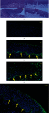
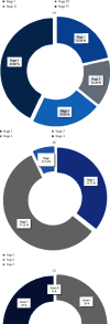
Methods: Fourteen eyes of 8 patients were included in the study. Foster's stage 1-4 OCP patients were given human intravenous immunoglobulin, whereas patients with active inflammation were treated with prednisone tablets and methotrexate. Those who were intolerant to methotrexate and had severe inflammatory symptoms were treated with cyclophosphamide. Cataract surgery was performed for all patients after three months of systemic treatment under stable conditions. The conjunctival biopsy was evaluated by immunofluorescence microscopy. Then, patients were divided into individuals with or without ankyloblepharon. Records were reviewed for OCP stage, type of surgery, best-corrected visual acuity (BCVA), Schirmer I test, corneal fluorescein sodium staining, meibomian gland coloboma range, and ocular surface disease index (OSDI) scores. Follow-up was for the duration of taking topical and systemic medication.
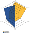
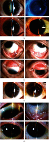
Results: Nine female (64.29%) and 4 male (35.71%) eyes were diagnosed with OCP by biopsy. The mean follow-up time was 60.64 ± 35.62 months. Thirteen eyes (92.86%) of 7 patients underwent phacoemulsification. One eye underwent phacoemulsification combined with amniotic membrane transplantation. The intracapsular extraction of cataract was applied to one eye. The BCVA improved significantly in all the patients, which remained stable until the last follow-up. The Schirmer I test was higher than that before the surgery. Corneal fluorescein sodium staining after surgery showed a decrease in score compared to the preoperative score. The BCVA of the patients after surgery increased significantly. The OSDI scores of patients with ankyloblepharon were significantly higher than for those without it. Postoperative symblepharon showed no significant difference compared to the preoperative symblepharon.
Conclusions: In this series, OCP patients with cataracts were able to undergo phacoemulsification surgery, whereas routine use of immunosuppression and closed postoperative follow-up were necessary.
Copyright © 2022 Yuan He et al.
Conflict of interest statement
The authors declare no competing interests.
Figures


References
-
- Chan L. S., Ahmed A. R., Anhalt G. J., et al. The first international consensus on mucous membrane pemphigoid: definition, diagnostic criteria, pathogenic factors, medical treatment, and prognostic indicators. Archives of Dermatology . 2002;138(3):370–379. doi: 10.1001/archderm.138.3.370. - DOI - PubMed
-
- Kirzhner M., Jakobiec F. A. Presented at Seminars in ophthalmology . Vol. 26. Taylor & Francis; 2011. Ocular cicatricial pemphigoid: a review of clinical features, immunopathology, differential diagnosis, and current management; pp. 270–277. - PubMed
Publication types
MeSH terms
Substances
LinkOut - more resources
Full Text Sources
Medical

