. 2022 Jul;149(8):1094-1105.
doi: 10.1017/S0031182022000622. Epub 2022 May 10.

New cytogenetic data on Caryophyllaeus laticeps and Paracaryophyllaeus gotoi, parasites of evolutionary interest

Affiliations

New cytogenetic data on Caryophyllaeus laticeps and Paracaryophyllaeus gotoi, parasites of evolutionary interest

Martina Orosová et al. Parasitology. 2022 Jul.

Abstract

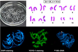
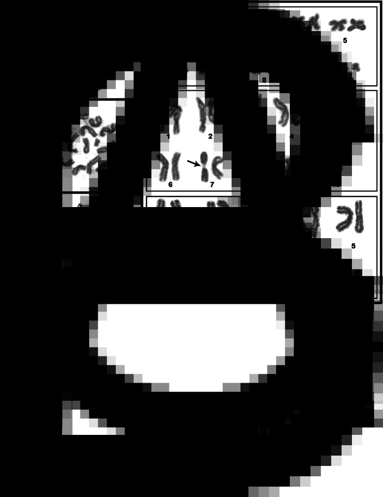
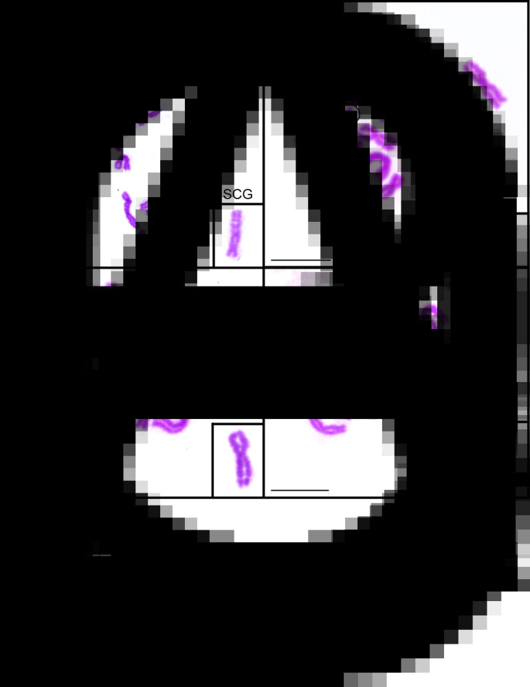
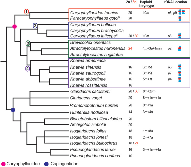
Caryophyllideans are intestinal parasites of freshwater fishes, occupying a basal position among the ‘true’ tapeworms. We performed detailed cytogenetic analyses of the well-known caryophyllidean species Caryophyllaeus laticeps. For comparison, we also examined for the first time the chromosomes of Paracaryophyllaeus gotoi, a specific parasite of loaches in China. Both species showed a diploid chromosome number of 2n = 20, n = 10m. Chromomycin A3 (CMA3)/diamidino-2-phenylindole (DAPI) staining performed for the first time in the class Cestoda revealed CMA3+/DAPI bands in the pericentromeric regions of the short arms of chromosome pair no. 7 in the karyotype of C. laticeps. Fluorescence in situ hybridization with the 18S rDNA probe confirmed the presence of a single cluster of major rDNA near the centromere on a pair of small chromosomes in both species. These findings support the hypothesis that the ancestral state in the family Caryophyllaeidae is a single interstitial cluster of major rDNA genes and thus one nucleolar organizer region per haploid genome. Our results, which we presented together with literature data plotted on a phylogenetic tree, show stability of caryophyllidean karyotypes at the genus level, but showed differences between genera without a clear phylogenetic signal. The data allowed us to at least formulate a hypothesis about the ancestral haploid chromosome number of n = 10 for the family Caryophyllaeidae and possibly for the sister family Capingentidae. In addition, we compared two populations of C. laticeps from water bodies with different levels of polychlorinated biphenyl contamination, showing a slightly increased incidence of chromosomal abnormalities at the contaminated site.

Keywords: Chromosome aberration; FISH; environmental pollution; karyotype; karyotype evolution; ribosomal DNA.


Conflict of interest statement

The authors declare there are no conflicts of interest.

Figures

None
Graphical abstract
Fig. 1.
Fig. 1.
Giemsa-stained chromosomes of Caryophyllaeus laticeps. (A–C) Mitotic chromosomes and karyotypes derived from mitotic metaphases, 2n = 20, n = 10 m. Arrows indicate NOR. Scale bar = 10 μm.
Fig. 2.
Fig. 2.
Silver nitrate-stained chromosomes of C. laticeps showing nucleoli and NOR location (arrows). (A) Interphase nucleus with one large nucleolus. (B) Pachytene nucleus with nucleolus residue on one chromosome bivalent. (C) Diplotene stage showing a single NOR site on the bivalent no. 7. (D) Metaphase II. (E) Anaphase II, two cells with a single NOR signal on four chromatids. (F) Haploid spermatid. Scale bar = 10 μm.
Fig. 3.
Fig. 3.
FISH with the 18S rDNA probe (red) on C. laticeps chromosomes. (A) Two mitotic metaphases; note a pair of chromosomes with clusters of 18S rDNA signals on each chromatid near the centromere (arrowhead). (B) Interphase nucleus. (C) Two leptotene nuclei. (D) Zygotene. (E) Pachytene bivalents showing a cluster of interstitial 18S rDNA signals associated with heterochromatic DAPI block. (F) Pachytene complement showing YOYO-1-stained nucleolar residues that co-localize with cluster of hybridization signals of the 18S rDNA probe. N, nucleolus. (G) Pachytene (stained only with DAPI) with distinct interstitial and terminal DAPI bands on the smallest bivalent (arrow). (H) Pachytene. (I) Diplotene, three cells. Asterisks indicate the seventh bivalent with rDNA cluster. (J) Eight metaphase II nuclei, each showing one chromosome with a cluster of interstitial 18S rDNA signals. (K) Graphical interpretation of metaphase II. (L) Early anaphase II with the smallest and biggest bivalents already separated (arrows). The chromosomes were counterstained with DAPI except for (F) (counterstained with YOYO-1). Scale bar = 10 μm.
Fig. 4.
Fig. 4.
Chromosomes of C. laticeps sequentially stained with CMA3/DAPI (A–F) and only with CMA3 (G–I). (A) Diplotene and (D) metaphase II after CMA3. (B) The same diplotene and (E) metaphase II after DAPI. (C, F) Merged images demonstrates that CMA3 positive region (red dot) is DAPI negative. In the small insets, the NOR-bearing bivalents showing CMA3+/DAPI bands in the pericentromeric region. (G) Pachytene nucleus with CMA3+ band (yellow). (H) Metaphase I nuclei showing chromosomes with GC-rich heterochromatic blocks. (I) Anaphase II nucleus. Arrows indicate the location of NOR (red dots). Scale bar = 10 μm.
Fig. 5.
Fig. 5.
Four types of CAs in mitotic metaphase cells of C. laticeps. (A) SCG; the inset shows enlarged SCG from a different metaphase. (B) CF; the inset shows enlarged CF from the same metaphase. (C) ISCG; the inset shows enlarged ISCG from a different metaphase. (D) SCB; the inset shows enlarged SCB from the same metaphase. Scale bar = 10 μm.
Fig. 6.
Fig. 6.
Chromosomes of Paracaryophyllaeus gotoi stained with AgNO3 (A–C) and FISH with the 18S rDNA probe (red) (D–I). (A, B) Pachytene cells with a large nucleolus. (C) Diakinesis. (D) Interphase nucleus. (E) Mitotic metaphase. (F, G) Pachytene bivalents showing a cluster of interstitial rDNA signals associated with a heterochromatic DAPI block of one bivalent. (H) Bouquet stage (zygotene) counterstained with YOYO-1; N, remnants of nucleolus. (I) Metaphase II. The chromosomes in (D–G) and (I) were counterstained with DAPI. Scale bar = 10 μm.
Fig. 7.
Fig. 7.
Overview of available data on chromosome number 2n/3n, chromosome morphology (a, acrocentric; m, metacentric; sm, submetacentric; t, telocentric; min, minute elements) and chromosomal pattern of rDNA in haploid genome (p, short chromosomal arm, q, long chromosomal arm) of caryophyllidean tapeworms along with their latest phylogenetic relationships based on Oros et al. (2018) and Scholz et al. (2011, 2015, 2021). *Data obtained in this study.

Similar articles

Cited by

References

    1. Bajer A (1959) Change of length and volume of mitotic chromosomes in living cells. Hereditas 45, 579–596.
    1. Barčák D, Oros M, Hanzelová V and Scholz T (2017) A synoptic review of Caryophyllaeus Gmelin, 1790 (Cestoda: Caryophyllidea), parasites of cyprinid fishes. Folia Parasitologica 64, 027. - PubMed
    1. Bazsalovicsová E, Kráľová-Hromadová I, Brabec J, Hanzelová V, Oros M and Scholz T (2014) Conflict between morphology and molecular data: a case of the genus Caryophyllaeus (Cestoda, Caryophyllidea), monozoic tapeworm of cyprinid fishes. Folia Parasitologica 61, 346–352. - PubMed
    1. Bombarová M and Špakulová M (2015) New chromosome characteristics of the monozoic tapeworm Caryophyllaeus laticeps (Cestoda, Caryophyllidea). Helminthologia 52, 336–340.
    1. Brázová T, Miklisová D, Barčák D, Uhrovič D, Šalamún P, Orosová M and Oros M (2021) Hazardous pollutants in the environment: fish host–parasite interactions and bioaccumulation of polychlorinated biphenyls. Environmental Pollution 291, 118175. - PubMed

Publication types