

Reflectance Confocal Microscopy and Electrical Impedance Spectroscopy in the Early Detection of Melanoma in Changing Lesions during Long-term Follow-up of Very High-risk Patients
- PMID: 35535641
- PMCID: PMC9558334
- DOI: 10.2340/actadv.v102.1105
Reflectance Confocal Microscopy and Electrical Impedance Spectroscopy in the Early Detection of Melanoma in Changing Lesions during Long-term Follow-up of Very High-risk Patients
Abstract
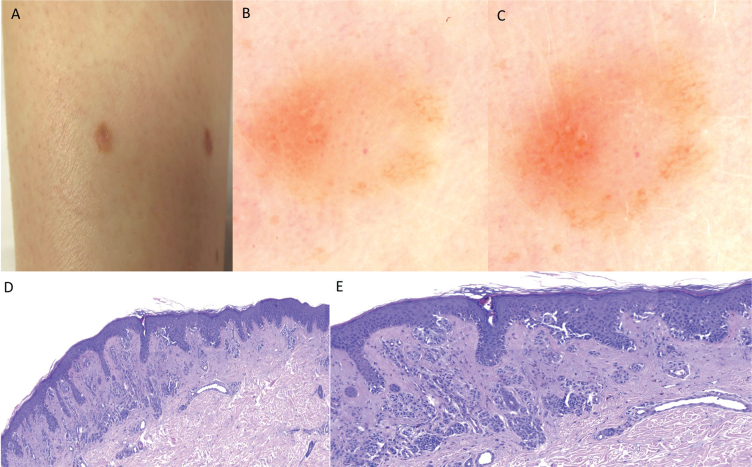
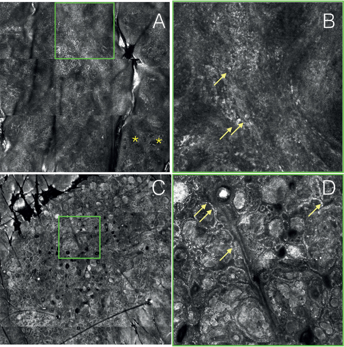
Electrical impedance spectroscopy has clinical relevance in diagnosing malignancy in melanocytic lesions. Sixty-eight lesions with changes during digital follow-up of patients at very high risk of developing melanoma were prospectively included in this study from February to December 2016. Electrical impedance spectroscopy and reflectance confocal microscopy were performed to evaluate their performance in this subset of difficult lesions. Forty-six lesions were considered suspicious on reflectance confocal microscopy and were excised, of these, 19 were diagnosed as melanoma. Fifteen melanomas were detected by electrical impedance spectroscopy, while 4 received a score lower than 4, which suggested no malignancy. The addition of reflectance confocal microscopy improves accuracy while maintaining the same sensitivity. In the case of electrical impedance spectroscopy scores <4, lesions exhibiting changes in follow-up may need short-term monitoring or excision if dermoscopy shows criteria for melanoma. Results of electrical impedance spectroscopy in this subset of very early lesions should be carefully considered due to the risk of false negatives.
Conflict of interest statement
Figures

References
-
- Malvehy J, Puig S. Follow-up of melanocytic skin lesions with digital total-body photography and digital dermoscopy: a two-step method. Clin Dermatol 2002; 20: 297–304. - PubMed
-
- Salerni G, Carrera C, Lovatto L, Puig-Butille J A, Badenas C, Plana E, et al. . Benefits of total body photography and digital dermatoscopy (“two-step method of digital follow-up”) in the early diagnosis of melanoma in patients at high risk for melanoma. J Am Acad Dermatol 2012; 67: e17–e27. - PMC - PubMed
-
- Moloney FJ, Guitera P, Coates E,Haass N, Ho K, Khoury R, et al. . Detection of primary melanoma in individuals at extreme high risk: a prospective 5-year follow-up study. JAMA Dermatol 2014; 150: 819–827. - PubMed
-
- Tromme I, Devleesschauwer B, Beutels P, Richez P, Praet N, Sacré L, et al. . Selective use of sequential digital dermoscopy imaging allows a cost reduction in the melanoma detection process: a Belgian study of patients with a single or a small number of atypical nevi. PLoS ONE 2014;9: e109339–e109339. - PMC - PubMed
-
- Lovatto L, Carrera C, Salerni G, Alós L, Malvehy J, Puig S. In vivo reflectance confocal microscopy of equivocal melanocytic lesions detected by digital dermoscopy follow-up. J Eur Acad Dermatology Venereol 2015; 29: 1918–1925. - PubMed
