

White matter microstructural variability linked to differential attentional skills and impulsive behavior in a pediatric population
- PMID: 35535719
- PMCID: PMC9977366
- DOI: 10.1093/cercor/bhac180
White matter microstructural variability linked to differential attentional skills and impulsive behavior in a pediatric population
Erratum in
-
Correction to: white matter microstructural variability linked to differential attentional skills and impulsive behavior in a pediatric population.Cereb Cortex. 2022 Oct 8;32(20):4640. doi: 10.1093/cercor/bhac367. Cereb Cortex. 2022. PMID: 36057836 Free PMC article. No abstract available.
Abstract
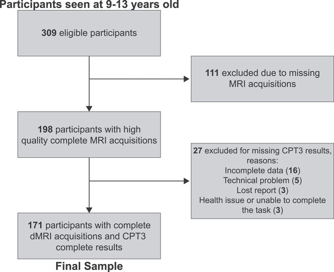
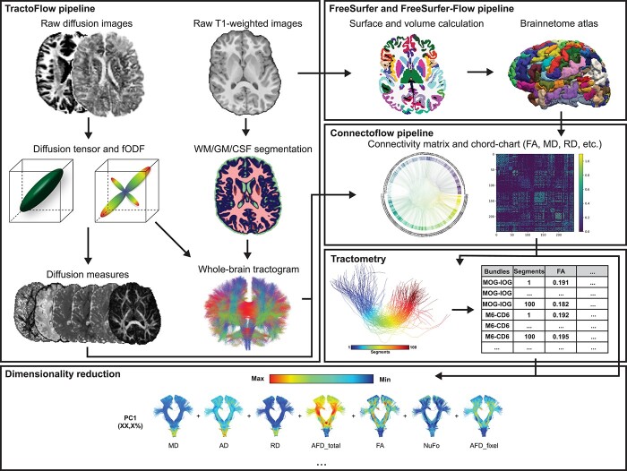
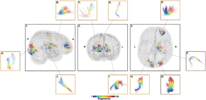
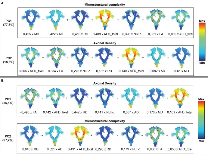
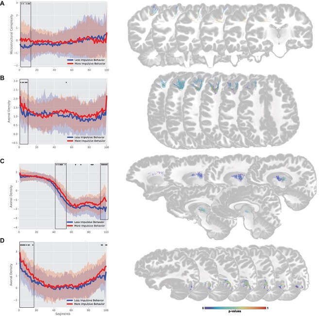
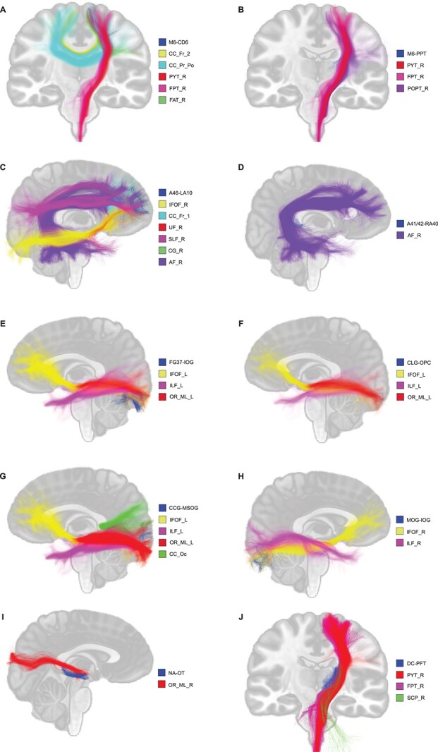
Structural and functional magnetic resonance imaging (MRI) studies have suggested a neuroanatomical basis that may underly attention-deficit-hyperactivity disorder (ADHD), but the anatomical ground truth remains unknown. In addition, the role of the white matter (WM) microstructure related to attention and impulsivity in a general pediatric population is still not well understood. Using a state-of-the-art structural connectivity pipeline based on the Brainnetome atlas extracting WM connections and its subsections, we applied dimensionality reduction techniques to obtain biologically interpretable WM measures. We selected the top 10 connections-of-interests (located in frontal, parietal, occipital, and basal ganglia regions) with robust anatomical and statistical criteria. We correlated WM measures with psychometric test metrics (Conner's Continuous Performance Test 3) in 171 children (27 Dx ADHD, 3Dx ASD, 9-13 years old) from the population-based GESTation and Environment cohort. We found that children with lower microstructural complexity and lower axonal density show a higher impulsive behavior on these connections. When segmenting each connection in subsections, we report WM alterations localized in one or both endpoints reflecting a specific localization of WM alterations along each connection. These results provide new insight in understanding the neurophysiology of attention and impulsivity in a general population.
Keywords: child & adolescent; cognitive functions; diffusion MRI; tractography; white matter connectivity.
© The Author(s) 2022. Published by Oxford University Press. All rights reserved. For permissions, please e-mail: journals.permissions@oup.com.
Figures
References
-
- Almeida LG, Ricardo-Garcell J, Prado H, Barajas L, Fernández-Bouzas A, Ávila D, Martínez RB. Reduced right frontal cortical thickness in children, adolescents and adults with ADHD and its correlation to clinical variables: a cross-sectional study. J Psychiatr Res. 2010:44(16):1214–1223. 10.1016/j.jpsychires.2010.04.026. - DOI - PubMed
-
- Ameis SH, Lerch JP, Taylor MJ, Lee W, Viviano JD, Pipitone J, Nazeri A, Croarkin PE, Voineskos AN, Lai M-C, et al. A diffusion tensor imaging study in children with ADHD, autism spectrum disorder, OCD, and matched controls: distinct and non-distinct white matter disruption and dimensional brain-behavior relationships. Am J Psychiatry. 2016:173(12):1213–1222. 10.1176/appi.ajp.2016.15111435. - DOI - PubMed
Publication types
MeSH terms
Grants and funding
LinkOut - more resources
Full Text Sources
Medical
