. 2022 Aug 25;40(8):736-750.
doi: 10.1093/stmcls/sxac033.

Krüppel-like Factor 4 Supports the Expansion of Leukemia Stem Cells in MLL-AF9-driven Acute Myeloid Leukemia

Affiliations

Krüppel-like Factor 4 Supports the Expansion of Leukemia Stem Cells in MLL-AF9-driven Acute Myeloid Leukemia

Andrew Henry Lewis et al. Stem Cells. .

Abstract

Acute myeloid leukemia (AML) is an aggressive malignancy of the bone marrow with 5-year overall survival of less than 10% in patients over the age of 65. Limited progress has been made in the patient outcome because of the inability to selectively eradicate the leukemic stem cells (LSC) driving the refractory and relapsed disease. Herein, we investigated the role of the reprogramming factor KLF4 in AML because of its critical role in the self-renewal and stemness of embryonic and cancer stem cells. Using a conditional Cre-lox Klf4 deletion system and the MLL-AF9 retroviral mouse model, we demonstrated that loss-of-KLF4 does not significantly affect the induction of leukemia but markedly decreased the frequency of LSCs evaluated in limiting-dose transplantation studies. Loss of KLF4 in leukemic granulocyte-macrophage progenitors (L-GMP), a population enriched for AML LSCs, showed lessened clonogenicity and percentage in the G2/M phase of the cell cycle. RNAseq analysis of purified L-GMPs revealed decreased expression of stemness genes and MLL-target genes and upregulation of the RNA sensing helicase DDX58. However, silencing of DDX58 in KLF4 knockout leukemia indicated that DDX58 is not mediating this phenotype. CRISPR/Cas9 deletion of KLF4 in MOLM13 cell line and AML patient-derived xenograft cells showed impaired expansion in vitro and in vivo associated with a defective G2/M checkpoint. Collectively, our data suggest a mechanism in which KLF4 promotes leukemia progression by establishing a gene expression profile in AML LSCs supporting cell division and stemness.

Keywords: KLF4; MLL-AF9; leukemic stem cells; transcription factor.


Figures

None
KLF4 promotes leukemic stem cell survival in MLL-AF9 acute myeloid leukemia by regulating genes in the leukemic stem cell signature, MLL targets, and cell cycle.
Figure 1.
Figure 1.
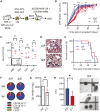
KLF4 is dispensable for the development of MLL-AF9-induced murine AML but supports disease progression. (A) Schematic diagram of MLL-AF9 (MA9)-induced leukemia using LSK cells from mice with conditional Klf4 deletion. (B) Flow cytometric detection of GFP+ CD11b+ leukemic cells in peripheral blood of fl/fl and Δ/Δ MA9 mice expressed as a percentage of total live cells (n = 10/group). (C) White blood cell counts at 5 weeks post-transplantation. (D) Representative blood smear of fl/fl and Δ/Δ MA9 mice at 7 weeks post-transplantation. (E) Kaplan–Meier analysis of survival of fl/fl and Δ/Δ MA9 mice (n = 10/group). (F) Frequency of myeloid (CD11b, Gr-1) and lymphoid (TCRβ, CD19) cells within GFP+ cells in the bone marrow and the spleen of moribund MA9 mice as determined by flow cytometry. A representative pie chart is shown. (G) Spleen weight of moribund fl/fl and Δ/Δ leukemic mice (n = 4/group). Mean and individual values are shown. (H) Colony-forming cell assay of fl/fl and Δ/Δ MA9 bone marrow cells from moribund mice (n = 7/group). The representative morphology of colonies is shown on the right. The data are represented as mean ± SD and are representative of 3 independent experiments. ns, not statistically significant. *P < .05, **P < .01, ***P < .001. Two-tailed Student’s t test was used in B, C, G, H. Log-rank test was used in E.
Figure 2.
Figure 2.
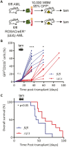
Post-transplant deletion of KLF4 delays leukemia progression. (A) Schematic diagram of induction of Klf4 deletion using the ROSA-CreER system. Cryopreserved leukemic cells were co-injected with whole bone marrow (WBM) in ablated mice. (B) Flow cytometric analysis of GFP+ CD11b+ cells in peripheral blood of fl/fl (n = 10) and iΔ/Δ (n = 11) MA9 mice after induction of gene deletion via daily administration of tamoxifen (Tam) beginning at day 14 post-transplantation and continuing for 5 days. (C) Kaplan–Meier analysis of survival of fl/fl (n = 10) and iΔ/Δ (n = 11) MA9 mice. *P < .05, ***P < .001. Two-tailed Student’s t test was used in B. Log-rank test was used in C.
Figure 3.
Figure 3.
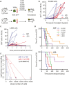
KLF4 regulates the frequency of MLL-AF9 leukemia-initiating cells. (A) Schematic diagram of limiting-dose transplantation to enumerate leukemia-initiating cell frequency. Leukemic bone marrow cells from 7 independent leukemic MA9 mice for each fl/fl and Δ/Δ group were injected, doses ranging from 1000 to 50 000, into sub-lethally irradiated mice (total of 35 mice per group). (B) Expansion of MA9 GFP+ cells in peripheral blood of mice transplanted with 50 000 fl/fl or Δ/Δ leukemic cells (n = 7/group). (C) Expansion of MA9 GFP+ cells in peripheral blood of mice transplanted with 1000 fl/fl or Δ/Δ leukemic cells (n = 7/group). (D) Overall survival of mice transplanted with limiting doses of fl/fl or Δ/Δ leukemic bone marrow cells (n = 7/cell dose). (E) Analysis using extreme limiting dilution analysis (ELDA) to determine the frequency of leukemia-initiating cells.
Figure 4.
Figure 4.
KLF4 knockout murine L-GMPs and human PDX cells show lower clonogenicity and impaired in vivo expansion. (A) Gating strategy to purify L-GMP cells from leukemic bone marrow by cell sorting. (B) Serial colony-forming cell assay in fl/fl or Δ/Δ MA9 L-GMP cells (n = 3/group, mean ± SD). (C) Freshly isolated (0 h) or in vitro cultured (48 h) fl/fl and Δ/Δ L-GMP cells were stained with propidium iodide for DNA content and cell cycle analysis (n = 3/group, mean ± SD). (D) Diagram of KLF4 gene deletion and transplantation in patient-derived xenograft (PDX) cells from an AML patient containing the MLL-AF9 rearrangement (Supplementary Fig. S5). (E) Genome editing of MLL-AF9 PDX cells with Cas9 only (control) or Cas9 and KLF4 sgRNA (KLF4 KO). Edited cells were transplanted into NSGS mice (n = 10/group) and monitored for the expansion of human CD45 leukemic cells in the blood. (F) Kaplan–Meier analysis of survival of mice transplanted with edited PDX cells (n = 10/group). *P < .05, ***P < .001. Two-tailed Student’s t test was used in B, C, and E. Log-rank test was used in F.
Figure 5.
Figure 5.
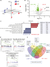
Loss-of-KLF4 results in loss of MLL targets and LSC gene signatures. (A) Principal component analysis of RNAseq performed in fl/fl and Δ/Δ L-GMP cells purified from 4 independent leukemic mice per group. (B) Volcano plot of deregulated genes in Δ/Δ L-GMP cells compared to fl/fl L-GMP cells (FDR<0.1). (C) Gene sets significantly enriched in fl/fl and Δ/Δ L-GMP with associated statistical values. (D) Selected GSEA enrichment plots. (E) Peacock plot comparing differentially expressed genes in fl/fl and Δ/Δ normal HSCs and L-GMP cells. Deregulated genes shared between human AML GMP cells and murine MA9 deregulated are indicated in linked boxes.
Figure 6.
Figure 6.
KLF4 suppression of DDX58 in MLL-AF9 L-GMP cells. (A) qPCR of DDX58 transcripts in fl/fl and Δ/Δ MA9 L-GMP cells purified from 4 independent leukemic mice per group. (B) Binding of endogenous KLF4 to the DDX58 promoter by ChIP-PCR. (C) Immunoblot detection of DDX58 protein in whole bone marrow (WBM) and L-GMP cells from fl/fl and Δ/Δ mice (n = 4/group). (D) Immunoblot analysis of the AKT-mTOR pathway in fl/fl and Δ/Δ L-GMP cells (n = 4/group). (E) Immunoblot of DDX58 in Δ/Δ MA9 scrambled shRNA (scr sh) and DDX58 shRNA (DDX58 sh) Δ/Δ MA9 cells. (F) The colony-forming ability of Δ/Δ MA9 scrambled shRNA (scr.sh) and DDX58 shRNA (DDX58 sh) cells in methylcellulose (n = 3). (G) Expansion of MA9 (Neptune+) and DDX58-shRNA (GFP+) or scramble-shRNA (GFP+) in peripheral blood post-transplantation (n = 10/group). *P < .05, *P < .01, ***P < .001. Two-tailed Student’s t test was used in A, B, F, and G.
Figure 7.
Figure 7.
Loss-of-KLF4 in MOLM13 impairs cell growth and G2/M cell cycle progression. (A) Confirmation of KLF4 deletion in MOLM13 and KLF4 KO clones via immunoblot. Cell growth of parental MOLM13 cells and 2 clones of KLF4 KO MOLM13 cells edited by CRISPR/Cas9 (mean ± SD). (B) Representative plots of cell cycle analysis by propidium iodide staining of MOLM13 and KLF4 KO cells following 24-h culture (n = 3). (C) Kaplan–Meier analysis of survival of NSG mice transplanted with MOLM13 and KLF4 KO cells (n = 6/group). (D) Heatmap of differentially expressed proteins in MOLM13 (parental) and 2 KLF4 KO cell clones detected by RPPA. (E) Immunoblot of MOLM13 KLF4 knockout detecting Cyclin B1 and DDX58. **P < .01, ***P < .001. Two-tailed Student’s t test was used in A and B. Log-rank test was used in C.

Similar articles

Cited by

References

    1. Madan V, Koeffler HP.. Differentiation therapy of myeloid leukemia: four decades of development. Haematologica. 2021;106(1):26-38. 10.3324/haematol.2020.262121. - DOI - PMC - PubMed
    1. Kantarjian H, Kadia T, DiNardo C, et al. . Acute myeloid leukemia: current progress and future directions. Blood Cancer J. 2021;11(2):41. 10.1038/s41408-021-00425-3. - DOI - PMC - PubMed
    1. Roboz GJ, Guzman M.. Acute myeloid leukemia stem cells: seek and destroy. Expert Rev Hematol. 2009;2(6):663-672. 10.1586/ehm.09.53. - DOI - PMC - PubMed
    1. Thomas D, Majeti R.. Biology and relevance of human acute myeloid leukemia stem cells. Blood. 2017;129(12):1577-1585. 10.1182/blood-2016-10-696054. - DOI - PMC - PubMed
    1. Shlush LI, Mitchell A, Heisler L, et al. . Tracing the origins of relapse in acute myeloid leukaemia to stem cells. Nature. 2017;547(7661):104-108. 10.1038/nature22993. - DOI - PubMed

Publication types

MeSH terms

Substances