Review
. 2022 Nov;17(11):2391-2398.
doi: 10.4103/1673-5374.338993.

Purinergic signaling systems across comparative models of spinal cord injury

Affiliations
Review

Purinergic signaling systems across comparative models of spinal cord injury

Eva E Stefanova et al. Neural Regen Res. 2022 Nov.

Erratum in

Abstract

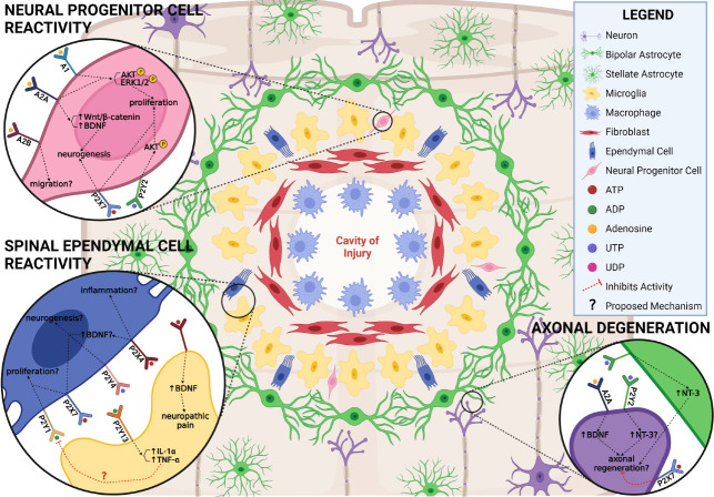
Within the last several decades, the scientific community has made substantial progress in elucidating the complex pathophysiology underlying spinal cord injury. However, despite the many advances using conventional mammalian models, both cellular and axonal regeneration following spinal cord injury have remained out of reach. In this sense, turning to non-mammalian, regenerative species presents a unique opportunity to identify pro-regenerative cues and characterize a spinal cord microenvironment permissive to re-growth. Among the signaling pathways hypothesized to be dysregulated during spinal cord injury is the purinergic signaling system. In addition to its well-known role as energy currency in cells, ATP and its metabolites are small molecule neurotransmitters that mediate many diverse cellular processes within the central nervous system. While our understanding of the roles of the purinergic system following spinal cord injury is limited, this signaling pathway has been implicated in all injury-induced secondary processes, including cellular death, inflammation, reactive gliosis, and neural regeneration. Given that the purinergic system is also evolutionarily conserved between mammalian and non-mammalian species, comparisons of these roles may provide important insights into conditions responsible for recovery success. Here, we compare the secondary processes between key model species and the influence of purinergic signaling in each context. As our understanding of this signaling system and pro-regenerative conditions continues to evolve, so does the potential for the development of novel therapeutic interventions for spinal cord injury.

Keywords: cell death; differenriation; glia; inflammation; neurogenesis; proliferation; purinergic signaling; reactive gliosis; regeneration; spinal cord injury; teleost.


Conflict of interest statement

Conflicts of interest: The authors declare no conflicts of interest.

Figures

Figure 1
Figure 1
Timeline depicting the secondary cellular injury response following mammalian spinal cord injury. The primary mechanical trauma is exacerbated by prolonged cell death, widespread inflammation, reactive gliosis, and axonal degeneration. These events prevent successful regeneration and limit sensorimotor recovery. Created with BioRender.com with permissions and publication license.
Figure 2
Figure 2
Timeline depicting the secondary cellular injury response following non-mammalian spinal cord injury. The primary mechanical trauma induces transient cell death, controlled inflammation, reactive gliosis, neurogenesis, and axonal regeneration. These events conclude within weeks and facilitate recovery and restoration of locomotor function in non-mammalian vertebrates. Created with BioRender.com with permissions and publication license.
Figure 3
Figure 3
Purinergic signaling within the spinal cord microenvironment during the early injury response. The first several days following mammalian SCI are characterized by widespread cell death, migration of various cell types to the lesion, inflammation, and reactive gliosis. Identified roles for purinergic receptors in these processes is summarized. Created with BioRender.com with permissions and publication license. ADP: adenosine diphosphate; ATP: adenosine triphosphate; CD39: cluster of differentiation 39; Ca2+: calcium; CREB: cAMP response element-binding protein; ERK1/2: extracellular signal-regulated kinases 1/2; IL: interleukin; NGF2: nerve growth factor-2; NLRP1/3: NLR family pyrin domain containing 1/3; NT3: neurotrophin-3; STAT3: signal transducer and activator of transcription 3; TNF-α: tumor necrosis factor alpha; UDP: uridine diphosphate; UTP: uridine triphosphate. Created with BioRender.com.
Figure 4
Figure 4
Purinergic signaling within the spinal cord microenvironment during the chronic injury response. After the first week following mammalian SCI, reactive astrocytes become scar forming, ependymal and neural progenitor cells fail to undergo injury-induced proliferation and neuronal differentiation, and axons continue to degenerate. Identified and hypothesized roles for purinergic receptors in these processes is summarized. Created with BioRender.com with publication permissions and publication license. ADP: Adenosine diphosphate; Akt: protein kinase B; ATP: adenosine triphosphate; BDNF: brain derived neurotrophic factor; CD39: cluster of differentiation 39; Ca2+: calcium; CREB: cAMP response element-binding protein; ERK1/2: extracellular signal-regulated kinases 1/2; IL: interleukin; NGF2: nerve growth factor-2; NLRP1/3: NLR family pyrin domain containing 1/3; NT3: neurotrophin-3; STAT3: signal transducer and activator of transcription 3; TNF-α: tumor necrosis factor alpha; UDP: uridine diphosphate; UTP: uridine triphosphate; Wnt: wingless-related intergration site. Created with BioRender.com.

Similar articles

Cited by

References

    1. Ali AAH, Abdel-Hafiz L, Tundo-Lavalle F, Hassan SA, von Gall C. P2Y2 deficiency impacts adult neurogenesis and related forebrain functions. FASEB J. 2021;35:e21546. - PubMed
    1. Anderson MA, Burda JE, Ren Y, Ao Y, O'Shea TM, Kawaguchi R, Coppola G, Khakh BS, Deming TJ, Sofroniew MV. Astrocyte scar formation aids central nervous system axon regeneration. Nature. 2016;532:195–200. - PMC - PubMed
    1. Arthur DB, Georgi S, Akassoglou K, Insel PA. Inhibition of apoptosis by P2Y2 receptor activation: novel pathways for neuronal survival. J Neurosci. 2006;26:3798–3804. - PMC - PubMed
    1. Barry D, McDermott K. Differentiation of radial glia from radial precursor cells and transformation into astrocytes in the developing rat spinal cord. Glia. 2005;50:187–197. - PubMed
    1. Becker CG, Becker T, Hugnot JP. The spinal ependymal zone as a source of endogenous repair cells across vertebrates. Prog Neurobiol. 2018;170:67–80. - PubMed