. 2022 Nov;17(11):2465-2471.
doi: 10.4103/1673-5374.339008.

Effects of progesterone on T-type-Ca2+-channel expression in Purkinje cells

Affiliations

Effects of progesterone on T-type-Ca2+-channel expression in Purkinje cells

Annika Eickhoff et al. Neural Regen Res. 2022 Nov.

Abstract

Plasticity of cerebellar Purkinje cells (PC) is influenced by progesterone via the classical progesterone receptors PR-A and PR-B by stimulating dendritogenesis, spinogenesis, and synaptogenesis in these cells. Dissociated PC cultures were used to analyze progesterone effects at a molecular level on the voltage-gated T-type-Ca2+-channels Cav3.1, Cav3.2, and Cav3.3 as they helped determine neuronal plasticity by regulating Ca2+-influx in neuronal cells. The results showed direct effects of progesterone on the mRNA expression of T-type-Ca2+-channels, as well as on the protein kinases A and C being involved in downstream signaling pathways that play an important role in neuronal plasticity. For the mRNA expression studies of T-type-Ca2+-channels and protein kinases of the signaling cascade, laser microdissection and purified PC cultures of different maturation stages were used. Immunohistochemical staining was also performed to characterize the localization of T-type-Ca2+-channels in PC. Experimental progesterone treatment was performed on the purified PC culture for 24 and 48 hours. Our results show that progesterone increases the expression of Cav3.1 and Cav3.3 and associated protein kinases A and C in PC at the mRNA level within 48 hours after treatment at latest. These effects extend the current knowledge of the function of progesterone in the central nervous system and provide an explanatory approach for its influence on neuronal plasticity.

Keywords: Cav3.1; Cav3.2; Cav3.3; Purkinje cells; T-type-Ca2+-channels; neuronal plasticity; progesterone; protein kinase A; protein kinase C; rat cerebellum.


Conflict of interest statement

None

Figures

Figure 1
Figure 1
Experimental timeline.
Figure 2
Figure 2
Expression of T-type-Ca2+-channels in p0 (A) and p9 (B) PC of the cerebellum and dissociated PC (C). (A, B) Immunostaining of cryosections (12 µm in thickness) of rat cerebellum with antibodies against calbindin (red) and Cav3.1 (a), Cav3.2 (b), and Cav3.3 (c) in green. Cell nuclei are counterstained with Hoechst 33342 in blue. Scale bars: 20 µm. (C) Immunostaining of cultured PC (p0 + 2div) with antibodies against calbindin in red and Cav3.1 (a), Cav3.2 (b), and Cav3.3 (c) in green. Cell nuclei are counterstained with Hoechst in blue. Scale bars: 5 µm. eGCL/iGCL: External/internal granular cell layer; MCL: molecular cell layer; p0: The day of birth; p9: the 9th postnatal day; PC: Purkinje cell; PCL: Purkinje cell layer.
Figure 3
Figure 3
Expression of T-type-Ca2+-channels in laser microdissected PC. (A) Methylene blue-stained cryosections (12 µm in thickness) of p9 and p30 rat cerebella. White arrows mark microdissected PC. Scale bars: 100 µm. (B) Analysis of purity of microdissected PC via qRT-PCR based on mRNA expression of PC-specific markers (Calbindin), other neural cells (NeuN, alpha GABAR-6, and Vglut1) and astrocytes (GFAP) at the 9th postnatal day (p9) and the 30th postnatal day (p30) with GAPDH as a reference gene. All values were added and the percentage of each marker was calculated by estimating the portion of the total; n = 3. (C) Relative mRNA expression of CACNA1G, CACNA1H, and CACNA1I in microdissected PC at p9 and p30 normalized to GAPDH. The analysis was performed by qRT-PCR. Data are shown as the mean ± SEM; n = 4; statistical analyses with unpaired two-tailed t-test; significance level: P(CACNA1G) = 0.0006, P(CACNA1H) = 0.0004, P(CACNA1I) = 0.0481. *P < 0.05, ***P < 0.001. n = numbers of repetition of the experiment. eGCL/iGCL: External/internal granular cell layer; GAPDH: Glyceraldehyde-3-phosphate dehydrogenase; GCL: granular cell layer; GFAP: glial fibrillary acidic protein; MCL: molecular cell layer; NeuN: neuronal nuclei; PC: Purkinje cell; PCL: Purkinje cell layer; qRT-PCR: quantitative reverse transcription-polymerase chain reaction.
Figure 4
Figure 4
Expression of PGR in PC (A, B) and the effect of progesterone on T-type-Ca2+-channel mRNA expression in dissociated PC cultures (C). (A) Analysis of PGR mRNA expression in dissociated PC culture p0 (0 hours), 1 div (24 hours) and 2 div (48 hours) by quantitative reverse transcription-polymerase chain reaction with GAPDH as reference genes. Data are provided as the mean ± SEM; statistical analyses with unpaired two-tailed t-test. Significance level, p0–1div: not significant, 1–2div: P = 0.0301; n = 3. (B) Immunohistochemistry of dissociated PC p0 + 2div, anti-PGR is shown in green, anti-calbindin in red, nuclear staining was done with Hoechst 33342 in blue. Scale bar: 10 µm. (C) After incubation of dissociated p0 PC with 10 nM progesterone for 24 and 48 hours, the relative mRNA expression pattern of CACNA1G, CACNA1H, and CACNA1I normalized to GAPDH was evaluated; n = 4. Data are provided as the mean ± SEM; unpaired two-tailed t-test was performed for statistical analyses. Significance level: P(CACNA1G 24 hours) = 0.0246, P(CACNA1G 48 hours) = 0.0115, P(CACNA1H 24 hours) = 0.0063, P(CACNA1H 48 hours) = 0.0001, P(CACNA1I 24 hours) = 0.0006, P(CACNA1I 48 hours) = 0.0089. *P < 0.05, **P < 0.01, ***P < 0.001. n = numbers of repetition of the experiment. GAPDH: Glyceraldehyde-3-phosphate dehydrogenase; PGR: classical progesterone receptors A and B; PC: Purkinje cell.
Figure 5
Figure 5
Effects of progesterone on the mRNA expression of protein kinases. Relative mRNA expression pattern of the protein kinases PRKACA, PKCA, PRKCB, PRKCE, and PRKCZ normalized against GAPDH in dissociated p0 PC-cultures after treatment with 10 nM progesterone for 24 and 48 hours compared with unstimulated controls (quantitative reverse transcription-polymerase chain reaction); n = 4. Data is shown as the mean ± SEM; statistical analyses with unpaired two-tailed t-test. Significance level: P(PRKACA 24 hours) = 0.0487, P(PRKACA 48 hours) = 0.021, P(PKCA 24 hours) = 0.0151, P(PKCA 48 hours) = 0.0001, P(PRKCB 24 hours) = 0.0343, P(PRKCB 48 hours) = 0.0294, P(PRKCE 24 hours) = 0.0004, P(PRKCE 48 hours) = 0.0006, P(PRKCZ 24 hours) = 0.0053, P(PRKCZ 48 hours) = 0.0036. n = numbers of repetition of the experiment. *P < 0.05, **P < 0.01, ***P < 0.001. GAPDH: Glyceraldehyde-3-phosphate dehydrogenase; PC: Purkinje cell; PGR: classical progesterone receptors A and B.
Figure 6
Figure 6
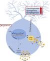
Schematic presentation of the hypothesis on the effects of progesterone on T-type-Ca2+-channels in Purkinje cells. Progesterone diffuses into PC where it influences gene expression in the nucleus via the classical PGR. It causes an increased expression of PKA and PKC, which leads to an increased Ca2+-influx. Due to increased expression of Cav3.1 and Cav3.3 induced by progesterone, Ca2+-influx is increased. More functional T-type-Ca2+-channels and an increased Ca2+-influx promote dendritogenesis, spinogenesis, and synaptogenesis, leading to neuronal plasticity. Ca2+: Calcium ions; PGR: classical progesterone receptors A and B; PKA: protein kinase A; PKC: protein kinase B.

Similar articles

Cited by

References

    1. Aguado C, García-Madrona S, Gil-Minguez M, Luján R. Ontogenic changes and differential localization of T-type Ca(2+) channel subunits Cav3.1 and Cav3.2 in mouse hippocampus and cerebellum. Front Neuroanat. 2016;10:83. - PMC - PubMed
    1. Arcos-Montoya D, Wegman-Ostrosky T, Mejía-Pérez S, De la Fuente-Granada M, Camacho-Arroyo I, García-Carrancá A, Velasco-Velázquez MA, Manjarrez-Marmolejo J, González-Arenas A. Progesterone receptor together with PKCαexpression as prognostic factors for astrocytomas malignancy. Onco Targets Ther. 2021;14:3757–3768. - PMC - PubMed
    1. Asmara H, Micu I, Rizwan AP, Sahu G, Simms BA, Zhang FX, Engbers JDT, Stys PK, Zamponi GW, Turner RW. A T-type channel-calmodulin complex triggers αCaMKII activation. Mol Brain. 2017;10:37. - PMC - PubMed
    1. Bannister RA, Beam KG. Ca(V)1.1: The atypical prototypical voltage-gated Ca²+channel. Biochim Biophys Acta. 2013;1828:1587–1597. - PMC - PubMed
    1. Baulieu EE. Neurosteroids: of the nervous system, by the nervous system for the nervous system. Recent Prog Horm Res. 1997;52:1–32. - PubMed