. 2022 Nov;17(11):2526-2529.
doi: 10.4103/1673-5374.339003.

In vivo neuronal and astrocytic activation in somatosensory cortex by acupuncture stimuli

Affiliations

In vivo neuronal and astrocytic activation in somatosensory cortex by acupuncture stimuli

Xiao-Yue Chang et al. Neural Regen Res. 2022 Nov.

Abstract

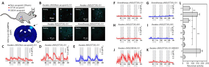
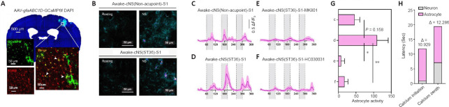
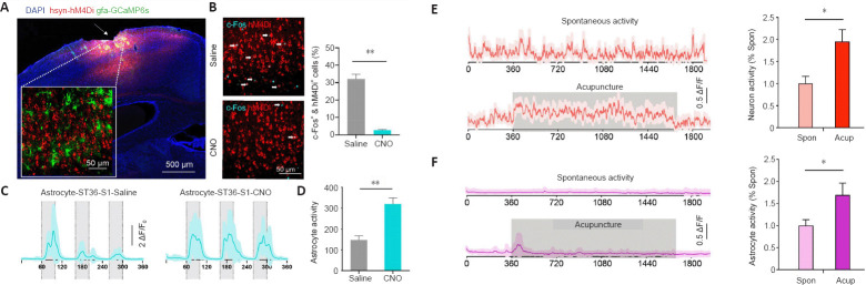
Acupuncture is a medical treatment that has been widely practiced in China for over 3000 years, yet the neural mechanisms of acupuncture are not fully understood. We hypothesized that neurons and astrocytes act independently and synergistically under acupuncture stimulation. To investigate this, we used two-photon in vivo calcium recording to observe the effects of acupuncture stimulation at ST36 (Zusanli) in mice. Acupuncture stimulation in peripheral acupoints potentiated calcium signals of pyramidal neurons and astrocytes in the somatosensory cortex and resulted in late-onset calcium transients in astrocytes. Chemogenetic inhibition of neurons augmented the astrocytic activity. These findings suggest that acupuncture activates neuronal and astrocytic activity in the somatosensory cortex and provide evidence for the involvement of both neurons and astrocytes in acupuncture treatment.

Keywords: N-methyl-D-aspartate receptor; acupuncture; astrocyte; chemogenetic; imaging; neuron; somatosensory cortex; transient receptor potential A1; two-photon in vivo.


Conflict of interest statement

Conflicts of interest: The authors declare no conflicting interests.

Figures

Figure 1
Figure 1
Study flowchart.
Figure 2
Figure 2
Acupuncture induces calcium transients in S1 neurons. (A) Top, schematic diagram showing the acupoints ST36 (Zusanli) and GB34 (Yanglingquan), and non-acupoint in adjacent sites as the sham control group; bottom, transfection sites (arrow) of GCaMP6s in S1. Scale bar: 500 μm. (B) Pseudo-colored images reflecting calcium intensity of neurons in selected field of view from S1 under resting state and during acupuncture. Scale bar: 50 μm. (C–K) Time-series records of normalized calcium values (in ΔF/F0) during the period of repeated acupuncture stimuli (grey shaded box). The temporal scale (x-axis) was presented in seconds. Three acupuncture courses (20 seconds each) were sequentially applied, with a 30-second resting interval between two treatments. In each group, calcium activities from representative fields of view were normalized and averaged for plotting. Fields of view numbers: n = 10 for C; n = 8 for D, K; n = 3 for E–G, J; n = 6 for H–I. Animal numbers: n = 3 each. (L) Comparison of total integrated neuronal calcium activity during the acupuncture course. Data are expressed as mean ± SEM and were analyzed by one-way analysis of variance. Group effect: F(8, 41) = 20.10, P < 0.001, Bonferroni’s multiple comparison test: **P < 0.01. cNS: Needle stimulation on the contralateral side; GCaMP6s: green fluorescent-calmodulin protein 6s; iNS: needle stimulation on the ipsilateral side; M1: primary motor cortex; NS: needle stimulation; S1: primary somatosensory cortex.
Figure 3
Figure 3
Acupuncture activates calcium transients in astrocytes. (A) Transfection sites of AAV-gfaABC1D-GCaMP6f in S1 were colocalized with astrocyte marker S100β (white arrowheads, red, stained by Alexa Fluor 594). Scale bars: 50 μm. (B) Pseudo-colored images for the intensity of astrocytic calcium spikes in selected fields of view during the resting state and NS. Scale bars: 50 μm. (C–F) Time-series recordings of normalized calcium values (in ΔF/F0) during the acupuncture sessions (grey shaded box). The temporal scale (x-axis) was presented in seconds. The protocol of acupuncture was the same as in Figure 2. In each group, the calcium activity from different fields of view was recorded for data plotting. Field of view numbers: n = 9 for C–D, n = 5 for E, n = 10 for F. Animal numbers: n = 3 each. (G) Comparison of total integrated astrocytic calcium activity. Data are expressed as mean ± SEM and were analyzed by one-way analysis of variance. Group effect: F(3, 29) = 5.188, P = 0.0054; Bonferroni’s multiple comparison test: *P < 0.05, **P < 0.01. (H) Comparison of kinetics of calcium spikes between neurons and astrocytes from the recordings under cNS stimulation on ST36 of awake mice. cNS: Needle stimulation on the contralateral side; DAPI: 4′,6-diamidino-2-phenylindole; NS: needle stimulation; S1: primary somatosensory cortex.
Figure 4
Figure 4
Astrocytic calcium activity is mediated by neurons. (A) Transfection of hM4Di into excitatory neurons of S1 (arrow) and transfection of gfaABC1D-GCaMP6s into astrocytes. Scale bar: 500 μm. (B) Co-staining (arrows) of c-Fos (blue) and hM4Di (red) in S1. Mice were administered CNO, followed by astrocytic calcium imaging as in C, and were immediately sacrificed for brain sectioning and immunofluorescence assay. Scale bar: 50 μm. Right, CNO treatment significantly suppressed the activity of transfected neurons (Mann-Whitney U test, **P = 0.0012). (C) Time-series recording of normalized astrocytic calcium signals during acupuncture treatment. Grey shades indicate the acupuncture period. Approximately 15–20 cells from four animals were plotted. (D) Total integrated calcium activity was potentiated by CNO treatment (Mann-Whitney U test, **P = 0.0022). (E) Left, time-series recordings of normalized neuronal calcium activity under resting state or during 20-minute continuous manual acupuncture on ST36. A total of 18 fields of view (FOVs) from four animals were plotted in each group. Right, the total neuronal calcium activity was elevated under acupuncture (Mann-Whitney U test, *P = 0.0293). (F) Left, time-series recordings of normalized astrocytic calcium activity under resting state or during the acupuncture on ST36. A total of 18 fields of view from four animals were plotted in each group. Right, the total astrocytic calcium activity was elevated during acupuncture (Mann-Whitney U test, *P = 0.0303). The temporal scale (x-axis) of c–f was presented in seconds. Data are expressed as mean ± SEM. The experiments were repeated twice. Acup: During the acupuncture on ST36; CNO: clozapine-N-oxide; DAPI: 4′,6-diamidino-2-phenylindole; GCaMP6s: green fluorescent-calmodulin protein 6s; S1: primary somatosensory cortex; Spon: resting state.

Similar articles

Cited by

References

    1. Allen NJ, Lyons DA. Glia as architects of central nervous system formation and function. Science. 2018;362:181–185. - PMC - PubMed
    1. Aronoff R, Matyas F, Mateo C, Ciron C, Schneider B, Petersen CC. Long-range connectivity of mouse primary somatosensory barrel cortex. Eur J Neurosci. 2010;31:2221–2233. - PubMed
    1. Brancaccio M, Patton AP, Chesham JE, Maywood ES, Hastings MH. Astrocytes control circadian timekeeping in the suprachiasmatic nucleus via glutamatergic signaling. Neuron. 2017;93:1420–1435. e5. - PMC - PubMed
    1. Bushnell MC, Duncan GH, Hofbauer RK, Ha B, Chen JI, Carrier B. Pain perception: is there a role for primary somatosensory cortex? Proc Natl Acad Sci U S A. 1999;96:7705–7709. - PMC - PubMed
    1. Cichon J, Blanck TJJ, Gan WB, Yang G. Activation of cortical somatostatin interneurons prevents the development of neuropathic pain. Nat Neurosci. 2017;20:1122–1132. - PMC - PubMed