. 2022 Jun 28;13(3):e0109922.
doi: 10.1128/mbio.01099-22. Epub 2022 May 10.

Human Metapneumovirus Phosphoprotein Independently Drives Phase Separation and Recruits Nucleoprotein to Liquid-Like Bodies

Affiliations

Human Metapneumovirus Phosphoprotein Independently Drives Phase Separation and Recruits Nucleoprotein to Liquid-Like Bodies

Kerri Beth Boggs et al. mBio. .

Abstract

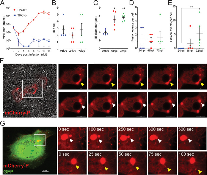
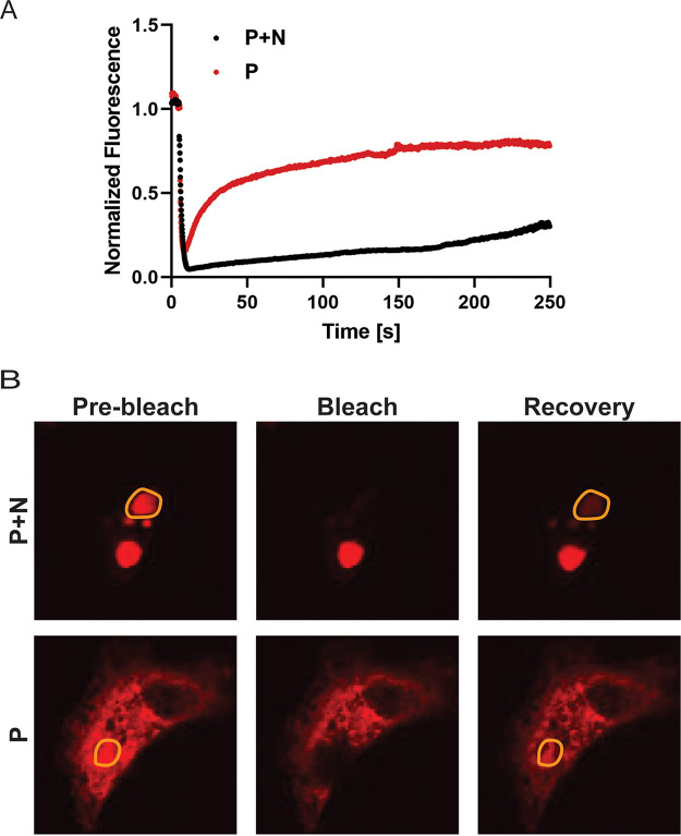
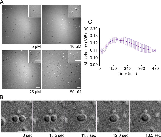
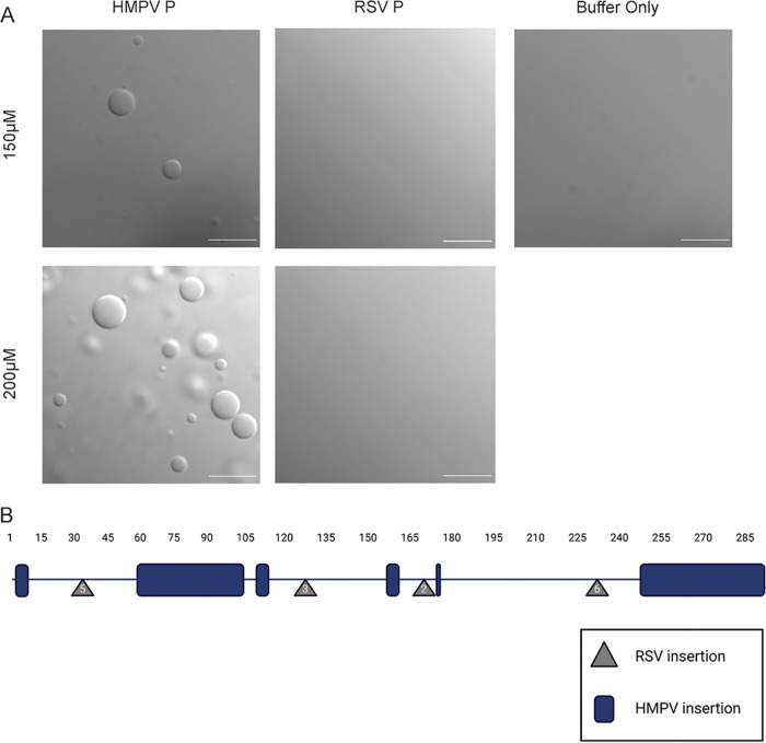
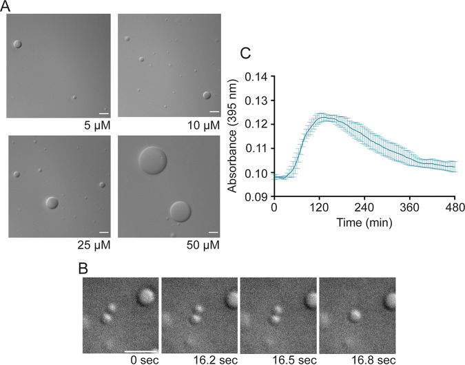
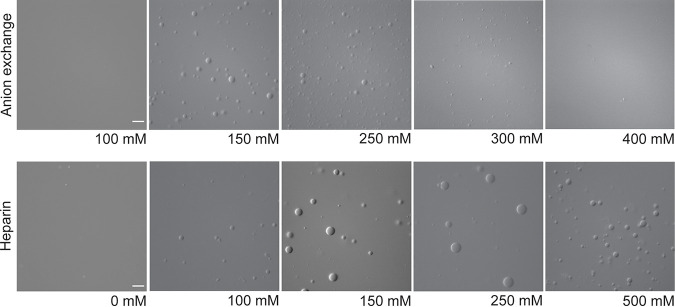
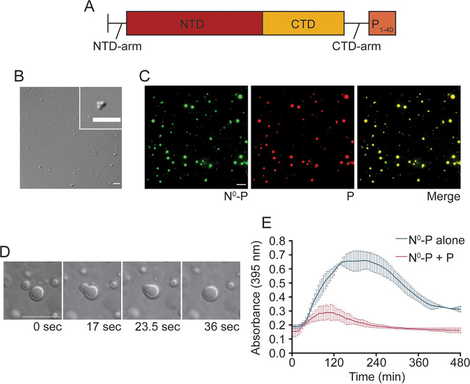
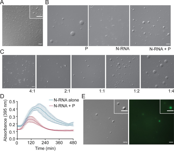
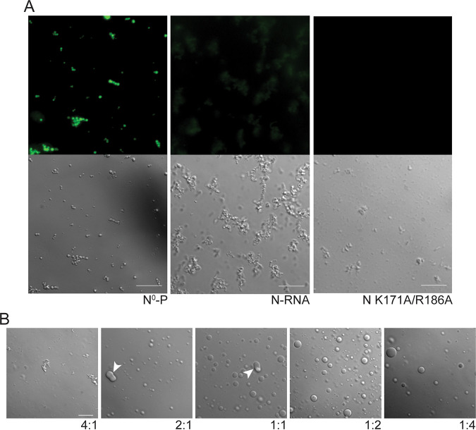
Human metapneumovirus (HMPV) inclusion bodies (IBs) are dynamic structures required for efficient viral replication and transcription. The minimum components needed to form IB-like structures in cells are the nucleoprotein (N) and the tetrameric phosphoprotein (P). HMPV P binds to the following two versions of the N protein in infected cells: N-terminal P residues interact with monomeric N (N0) to maintain a pool of protein to encapsidate new RNA and C-terminal P residues interact with oligomeric, RNA-bound N (N-RNA). Recent work on other negative-strand viruses has suggested that IBs are, at least in part, liquid-like phase-separated membraneless organelles. Here, HMPV IBs in infected or transfected cells were shown to possess liquid organelle properties, such as fusion and fission. Recombinant versions of HMPV N and P proteins were purified to analyze the interactions required to drive phase separation in vitro. Purified HMPV P was shown to form liquid droplets in isolation. This observation is distinct from other viral systems that also form IBs. Partial removal of nucleic acid from purified P altered phase-separation dynamics, suggesting that nucleic acid interactions play a role in IB formation. HMPV P also recruits monomeric N (N0-P) and N-RNA to droplets in vitro. These findings suggest that HMPV P may also act as a scaffold protein to mediate multivalent interactions with monomeric and oligomeric N, as well as RNA, to promote phase separation of IBs. Together, these findings highlight an additional layer of regulation in HMPV replication by the viral P and N proteins. IMPORTANCE Human metapneumovirus (HMPV) is a leading cause of respiratory disease among children, immunocompromised individuals, and the elderly. Currently, no vaccines or antivirals are available for the treatment of HMPV infections. Cytoplasmic inclusion bodies (IBs), where HMPV replication and transcription occur, represent a promising target for the development of novel antivirals. The HMPV nucleoprotein (N) and phosphoprotein (P) are the minimal components needed for IB formation in eukaryotic cells. However, interactions that regulate the formation of these dynamic structures are poorly understood. Here, we showed that HMPV IBs possess the properties of liquid organelles and that purified HMPV P phase separates independently in vitro. Our work suggests that HMPV P phase-separation dynamics are altered by nucleic acid. We provide strong evidence that, unlike results reported from other viral systems, HMPV P alone can serve as a scaffold for multivalent interactions with monomeric (N0) and oligomeric (N-RNA) HMPV N for IB formation.

Keywords: HMPV; inclusion bodies; phase separation; pneumovirus.


Conflict of interest statement

The authors declare no conflict of interest.

Figures

FIG 1
FIG 1
Characterization of a recombinant HMPV-mCherryP virus. (A) Vero cells were infected with an MOI of 0.1, and cells were kept in the absence (TPCK−) or presence (TPCK+) of trypsin until day 16 postinfection. Virus was harvested from the cell supernatants every other day and titrated. Vero cells were infected with HMPV-mCherryP virus using an MOI of 3 to quantify the number of IBs per cell (B) and IB diameter (C) at different times postinfection. Fusion (D) and fission (E) events were counted in Vero cells infected with HMPV-mCherryP at an MOI of 3, during a lapse of 10 min. Images were acquired every 30 sec using a LionHeartFX fluorescence microscope. (F) Time-lapse microscopy of Vero cells infected with HMPV-mCherryP, highlighting fission events (top; yellow arrowheads) and fusion events (bottom; white arrowheads). (G) Vero cells were electroporated with a plasmid encoding mCherryP and subsequently infected with rgHMPV-GFP virus. Forty-eight hours postinfection time-lapse microscopy was performed using a NikonA1 confocal microscope, with images acquired every 25 sec. Fusion (white arrowheads) and fission (yellow arrowheads) events are shown. Statistical analysis was performed using Student’s t test. *, P < 0.1; **, P < 0.01.
FIG 2
FIG 2
FRAP analysis of HMPV P protein in inclusion bodies and the cytosol. (A) Vero cells were transfected with pCAGGS plasmid expressing mCherry-P only (P) or cotransfected with pCAGGS plasmids encoding mCherry-P and N protein (P+N). At 24 h posttransfection, live-cell confocal microscopy was used to perform FRAP at 37°C on punctate regions by drawing a region of interest (ROI) representing a whole inclusion body or an equivalent area in the cytosol with P protein only. FRAP data were corrected for background, were normalized, and are represented as means from the recovery curves. (B) Live-cell confocal images collected during FRAP, showing recovery profiles of inclusions 4 min postbleaching. Bleaching was performed at 100% laser power.
FIG 3
FIG 3
Anion exchange-purified HMPV P phase separates independently in vitro. (A) Anion exchange-purified HMPV P was tested at concentrations ranging from 5 μM to 50 μM in a droplet assay (maximum droplet size, 3.4 μm). DIC microscopy imaging of droplets was performed with a 60× objective on a Nikon Eclipse E600 microscope. The scale bar is 10 μm. (B) Time lapse imaging of anion exchange purified HMPV P (80 μM) droplet fusion was acquired using a 100× oil objective on a Zeiss Axiovert 200M microscope. Scale bar, 5 μm. (C) Anion exchange-purified HMPV P (40 μM) was mixed with turbidity assay buffer in a clear 96-well plate. The solution was analyzed using a SpectraMax iD3 instrument to measure the absorbance at 395 nm at 5-min intervals with mixing.
FIG 4
FIG 4
RSV phosphoprotein alone is unable to undergo liquid-liquid phase separation. (A) HMPV P protein was tested parallel with purified RSV A2 P protein, using the same dextran buffer used to mimic the cytosol. RSV P is unable to undergo liquid-liquid phase separation in this study. Droplet assays were performed side by side. DIC microscopy was performed on the Axiovert 200M instrument with a 63× oil objective; scale bar, 10 μm. (B) A schematic of the HMPV P protein amino acid sequence, with representation of residue insertions within HMPV (blue), compared with RSV P, and RSV P insertions (gray), compared with HMPV P. HMPV P (HMPV; Uniprot Q8B9Q8) and RSV (RSV A2; Uniprot P03421) sequences were acquired through the Uniprot database and aligned using the NCBI alignment tool, with adjustments made based on observations in Cardone et al. (45). Made with BioRender.com.
FIG 5
FIG 5
Heparin-purified HMPV P phase separates independently in vitro. Heparin-purified HMPV P was tested at concentrations ranging from 5 μM to 50 μM in a droplet assay (maximum droplet size, >50 μm). DIC microscopy imaging of droplets was performed with a 60× objective on a Nikon Eclipse E600 microscope. Scale bar, 10 μm. (B) Time-lapse imaging of heparin-purified HMPV P (150 μM) droplet fusion was acquired using a 100× oil objective on a Zeiss Axiovert 200M microscope. Scale bar, 5 μm. (C) Heparin-purified HMPV P (40 μM) was mixed with turbidity assay buffer in a clear 96-well plate. The solution was analyzed using a SpectraMax iD3 instrument to measure the absorbance at 395 nm at 5-min intervals with mixing.
FIG 6
FIG 6
The presence of nucleic acid modulates HMPV P phase-separation dynamics. Anion exchange-purified HMPV P (15 μM) and heparin-purified HMPV P (15 μM) were tested in a droplet assay using buffers with different concentrations of KCl ranging from 0 mM to 500 mM. The DIC microscopy imaging of droplets was performed with a 60× objective on a Nikon Eclipse E600 microscope. Scale bar, 10 μm; the magnification is the same for all images.
FIG 7
FIG 7
HMPV P recruits N0-P to liquid droplets. (A) Schematic of the N0-P construct which includes full-length HMPV N fused to the first 40 amino acids of HMPV P. (B) HMPV N0-P (15 μM) was tested in a droplet assay. DIC images were acquired at different time points using a 60× objective on a Nikon Eclipse E600 instrument. Scale bar, 7 μm. (C) HMPV N0-P (15 μM) labeled with Alexa 488 TFP ester was mixed with anion exchange-purified HMPV P (15 μM) labeled with Alexa 594 NHS ester in a droplet assay. Fluorescence images were acquired using a 60× objective on a Nikon Eclipse E600 instrument. Scale bar, 10 μm. (D) HMPV N0-P (50 μM) was mixed with anion exchange-purified HMPV P (50 μM). Time-lapse imaging of N0-P/P droplet fusion was acquired using a 100× oil objective on a Zeiss Axiovert 200M microscope. Scale bar, 10 μm. (E) HMPV N0-P (40 μM) was tested alone or with anion exchange-purified HMPV P (40 μM) in a turbidity assay. The protein solutions were plated in a clear 96-well plate with turbidity assay buffer, and the absorbance was measured at 395 nm by a SpectraMax iD3 instrument at 5-min intervals.
FIG 8
FIG 8
HMPV P recruits N-RNA rings to liquid droplets. (A) HMPV N-RNA (25 μM) was tested in a droplet assay. The DIC microscopy imaging of droplets was performed with a 60× objective on a Nikon Eclipse E600 microscope. Scale bar, 10 μm. (B) HMPV N-RNA (15 μM) was mixed with heparin-purified HMPV P (15 μM) in a droplet assay. DIC images were acquired using a 60× objective on a Nikon Eclipse E600 instrument. Scale bar, 10 μm. (C) HMPV N-RNA and heparin-purified HMPV P were tested in a droplet assay at different ratios (4:1 for 20 μM N-RNA: 5 μM P; 2:1 for 10 μM N-RNA: 5 μM P; 1:1 for 5 μM N-RNA: 5 μM P; 1:2 for 5 μM N-RNA: 10 μM P; 1:4 for 5 μM N-RNA: 20 μM P). The DIC microscopy imaging of droplets was performed as described above. (D) HMPV N-RNA (40 μM) was tested alone or with heparin-purified HMPV P (40 μM) in a turbidity assay. The protein solutions were plated in a clear 96-well plate with turbidity assay buffer, and the absorbance was measured at 395 nm by a SpectraMax iD3 instrument at 5-min intervals with mixing. (E) HMPV N-RNA (15 μM), heparin-purified HMPV P (15 μM) and an RNA decamer tagged with 6-carboxyfluorescein on the 3′ end (5 μM) were mixed and tested in a droplet assay. The DIC and fluorescence microscopy imaging of droplets were performed as described above. Scale bar, 10 μm.
FIG 9
FIG 9
RNA-binding mutant HMPV N K171A/R186A forms gel-like droplets with P. (A) HMPV N0-P, N-RNA, or N K171A/R186A (15 μM) were tested in a droplet assay with an RNA decamer tagged with 6-carboxyfluorescein on the 3′ end (5 μM). DIC and fluorescence microscopy imaging were performed on a Zeiss Axiovert 200M instrument with a 63× oil objective. Scale bar, 10 μm. (B) HMPV N K171A/R186A and heparin-purified HMPV P were tested in a droplet assay at different ratios (4:1 for 20 μM N K171A/R186A: 5 μM P; 2:1 for 10 μM N K171A/R186A: 5 μM P; 1:1 for 5 μM N K171A/R186A: 5 μM P; 1:2 for 5 μM N K171A/R186A: 10 μM P; 1:4 for 5 μM N K171S/R186A: 20 μM P). The DIC microscopy imaging of droplets was performed on a Zeiss Axiovert 200M microscope with a 63× oil objective. White arrowheads indicate altered droplet fusion. Scale bar, 10 μm.

Similar articles

Cited by

References

    1. van den Hoogen BG, de Jong JC, Groen J, Kuiken T, de Groot R, Fouchier RA, Osterhaus AD. 2001. A newly discovered human pneumovirus isolated from young children with respiratory tract disease. Nat Med 7:719–724. doi:10.1038/89098. - DOI - PMC - PubMed
    1. Edwards KM, Zhu Y, Griffin MR, Weinberg GA, Hall CB, Szilagyi PG, Staat MA, Iwane M, Prill MM, Williams JV, New Vaccine Surveillance Network . 2013. Burden of human metapneumovirus infection in young children. N Engl J Med 368:633–643. doi:10.1056/NEJMoa1204630. - DOI - PMC - PubMed
    1. Williams JV, Harris PA, Tollefson SJ, Halburnt-Rush LL, Pingsterhaus JM, Edwards KM, Wright PF, Crowe JE, Jr.. 2004. Human metapneumovirus and lower respiratory tract disease in otherwise healthy infants and children. N Engl J Med 350:443–450. doi:10.1056/NEJMoa025472. - DOI - PMC - PubMed
    1. Schildgen V, van den Hoogen B, Fouchier R, Tripp RA, Alvarez R, Manoha C, Williams J, Schildgen O. 2011. Human metapneumovirus: lessons learned over the first decade. Clin Microbiol Rev 24:734–754. doi:10.1128/CMR.00015-11. - DOI - PMC - PubMed
    1. Rima B, Collins P, Easton A, Fouchier R, Kurath G, Lamb RA, Lee B, Maisner A, Rota P, Wang L, ICTV Report Consortium . 2017. ICTV virus taxonomy profile: Pneumoviridae. J Gen Virol 98:2912–2913. doi:10.1099/jgv.0.000959. - DOI - PMC - PubMed

Publication types

LinkOut - more resources