. 2022 Jun 30;28(7):gaac016.
doi: 10.1093/molehr/gaac016.

Uterine-specific SIRT1 deficiency confers premature uterine aging and impairs invasion and spacing of blastocyst, and stromal cell decidualization, in mice

Affiliations

Uterine-specific SIRT1 deficiency confers premature uterine aging and impairs invasion and spacing of blastocyst, and stromal cell decidualization, in mice

Magdalina J Cummings et al. Mol Hum Reprod. .

Abstract

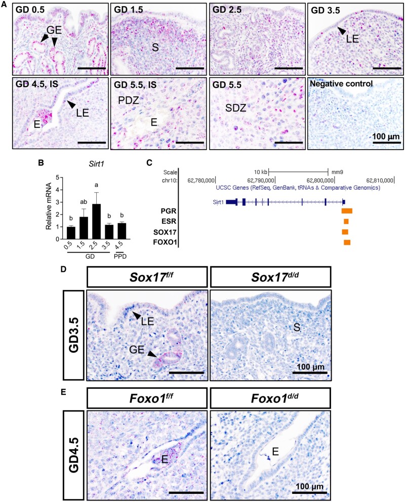
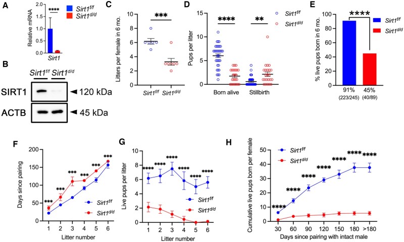
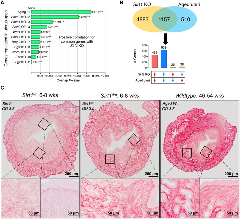
A distinct age-related alteration in the uterine environment has recently been identified as a prevalent cause of the reproductive decline in older female mice. However, the molecular mechanisms that underlie age-associated uterine adaptability to pregnancy are not known. Sirtuin 1 (SIRT1), a multifunctional NAD+-dependent deacetylase that regulates cell viability, senescence and inflammation during aging, is reduced in aged decidua. Thus, we hypothesize that SIRT1 plays a critical role in uterine adaptability to pregnancy and that uterine-specific ablation of Sirt1 gene accelerates premature uterine aging. Female mice with uterine ablation of Sirt1 gene using progesterone receptor Cre (PgrCre) exhibit subfertility and signs of premature uterine aging. These Sirt1-deficient mothers showed decreases in litter size from their 1st pregnancy and became sterile (25.1 ± 2.5 weeks of age) after giving birth to the third litter. We report that uterine-specific Sirt1 deficiency impairs invasion and spacing of blastocysts, and stromal cell decidualization, leading to abnormal placentation. We found that these problems traced back to the very early stages of hormonal priming of the uterus. During the window of receptivity, Sirt1 deficiency compromises uterine epithelial-stromal crosstalk, whereby estrogen, progesterone and Indian hedgehog signaling pathways are dysregulated, hampering stromal cell priming for decidualization. Uterine transcriptomic analyses also link these causes to perturbations of histone proteins and epigenetic modifiers, as well as adrenomedullin signaling, hyaluronic acid metabolism, and cell senescence. Strikingly, our results also identified genes with significant overlaps with the transcriptome of uteri from aged mice and transcriptomes related to master regulators of decidualization (e.g. Foxo1, Wnt4, Sox17, Bmp2, Egfr and Nr2f2). Our results also implicate accelerated deposition of aging-related fibrillar Type I and III collagens in Sirt1-deficient uteri. Collectively, SIRT1 is an important age-related regulator of invasion and spacing of blastocysts, as well as decidualization of stromal cells.

Keywords: SIRT1; Sirtuin 1; implantation; pregnancy; progesterone receptor signaling; stromal cell decidualization.


Conflict of interest statement

All authors declare no conflict of interest.

Figures

Figure 1.
Figure 1.
Advanced maternal age in mice impacts artificial decidua formation and Sirt1 mRNA expression. (A) Artificial decidua formation in uteri of young and aged C57BL/6 females at DD2 and DD5. Blue-dashed squares denote decidual tissue in the uterine horn. (B) Ratios of decidual uterine horn weight to control uterine horn weight in young and aged females at DD2 and DD5. (C) Histological analyses following H&E staining of uterine decidua in young and aged females at DD2 and DD5. (D–F) RNAscope in situ hybridization (D, F) and qRT-PCR analyses (E) of Sirt1 mRNA in decidua in young and aged females at DD2 and/or DD5. M, mesometrial side; AM, anti-mesometrial side. n = 6 for DD2; and n = 4 for DD5. DD2, decidual day 2; DD5, decidual day 5; H&E, hematoxylin and eosin; qRT-PCR, quantitative RT-PCR; Sirt1, Sirtuin 1. **P < 0.01; ***P < 0.001 (two-tailed Student’s t test). Data are presented as mean ± SEM. Also see Supplementary Figs. S1 and S2.
Figure 2.
Figure 2.
Regulation of expression of Sirt1 mRNA in uteri of young female mice during the peri-implantation period of pregnancy. (A) RNAscope in situ hybridization for Sirt1 mRNA in uteri of young female mice between GD 0.5 and 5.5 (n = 3). (B) Quantification of Sirt1 mRNA in uteri from young female mice between GD 0.5 and 3.5, as well as PPD 4.5 (n = 6). (C) Genome Browser tracks of PGR, ESR, SOX17 and FOXO1 binding at promoter region for Sirt1 gene in P4-treated, E2-treated, GD 3.5 or GD 4.5 uteri. UCSC Genome Browser views showing the mapped read coverage of PGR, ESR, SOX17 and FOXO1 ChIP-seq data (GSE34927. GSE36455, GSE118327 and GSE72892). (D) RNAscope in situ hybridization for Sirt1 mRNA in uteri from Sox17f/f and Sox17d/d mice (n = 3) on GD 3.5. (E) RNAscope in situ hybridization for Sirt1 mRNA in uteri from Foxo1f/f and Foxo1d/d mice (n = 3) on GD 4.5. E, embryo; GD, gestational day; GE, glandular epithelium; IIS, inter-implantation site; IS, implantation site; LE, luminal epithelium; PDZ, primary decidual zone; PPD, pseudopregnant day; S, stroma; SDZ, secondary decidual zone; Sirt1, Sirtuin 1. Different superscript letters denote significant differences (P < 0.05, ANOVA with Fisher’s LSD post-hoc test). Data are presented as mean ± SEM.
Figure 3.
Figure 3.
Uterine-specific deletion of Sirt1 impacts the fertility of female mice in a 6-month breeding trial. (A, B) Generation of Sirt1 conditional knockout (Sirt1d/d; PgrCre/+Sirt1f/f) female mice using the PgrCre mouse model was validated by using qRT-PCR (A) and western blot (B) analyses. Gene expression was normalized to 18s rRNA in qRT-PCR analyses; and β-actin (ACTB) was used as protein loading control in western blot analyses. ****P < 0.0001 (two-tailed Student’s t test). (C) Litters produced from Sirt1f/f (control) and Sirt1d/d female mice. Each dot represents the value for a different individual. ***P < 0.001(two-tailed Student’s t test). (D) Stillbirth and live pups born per litter in Sirt1f/f and Sirt1d/d female mice. Each dot represents the value for a different litter. **P < 0.01; ****P < 0.0001 (two-tailed Student’s t test). (E) Percent live pups born in 6 months from Sirt1f/f and Sirt1d/d female mice. ****P < 0.0001 (χ2 test). (F) Time taken to produce a specific number of litters. (G) Live pups born in a specific number of litters. (H) Cumulative live pups born since pairing. The mean number of pups produced up to each time point is denoted along with the respective error bar, with >180 days representing the day for calculating the total number of live pups produced. ***P < 0.001; ****P < 0.0001 (two-way ANOVA with Tukey’s multiple comparisons test) at specific time points for contrast between Sirt1f/f (in blue, n = 6) and Sirt1d/d (in red, n = 7) female mice. Data are presented as mean ± SEM. Also see Supplementary Figs. S3 and S4. The uncropped western blot is shown in Supplementary Fig. S3. Sirt1, Sirtuin 1; qRT-PCR, quantitative RT-PCR.
Figure 4.
Figure 4.
Uterine-specific deletion of Sirt1 in mice impacts spacing and invasion of blastocysts and stromal cell decidualization. (A, B) Images (A) and quantification (B) of implantation sites in Sirt1f/f and Sirt1d/d female mice (n = 6). NS, not significant (two-tailed Student’s t test). (C) Illustration of quantitation method for spacing of blastocysts within uterine horns. (D) Quantification of spacing of blastocysts in uteri from Sirt1f/f and Sirt1d/d females using the method depicted in (C). n = 6 females per groups. *P < 0.05 (two-tailed Student’s t test). (E) Decidual weight in Sirt1f/f and Sirt1d/d female mice (n = 6). ****P < 0.0001 (two-tailed Student’s t test). (F) Histological analyses following H&E staining of uteri from Sirt1f/f and Sirt1d/d female mice (n = 6). Dotted lines in GD 5.5 and 6.5 indicate the distance from implantation site to AM or M; and dotted lines in GD 9.5 indicate boundary between the fetal trophoblast compartment and the maternal decidua. M, mesometrial pole; AM, anti-mesometrial pole. (G) Ratio of the distance between E-to-M to distance between E-to-AM in uteri from Sirt1f/f and Sirt1d/d female mice at GD 5.5 and 6.5 (n = 6). E, embryo; Sirt1, Sirtuin 1. *P < 0.05 (two-tailed Student’s t test). (H) Artificial decidua formation and ratio of decidual to control horn weight in Sirt1f/f and Sirt1d/d female mice (n = 6). *P < 0.05 (two-tailed Student’s t test). Data are presented as mean ± SEM.
Figure 5.
Figure 5.
Immunohistochemical staining of proteins in uteri from Sirt1f/f and Sirt1d/d female mice on GD 5.5. PTGS2 (A), FOXO1 (B) and PGR (C). Black-dotted circles indicate sites of blastocyst implantation. Red-dotted lines indicate the boundary between the DSC and undifferentiated S. Inserts indicate high magnification of box area (n = 3). AM, anti-mesometrial pole; DSC, decidualized stromal cells; E, blastocyst; LE, luminal epithelium; M, mesometrial pole; S, stroma; Sirt1, Sirtuin 1.
Figure 6.
Figure 6.
Dysregulated estrogen and progesterone signaling in Sirt1-deficient mouse uteri during the window of receptivity to implantation by blastocysts. (A) Immunohistochemical staining of ESR1 and PGR in GD 3.5 uteri from Sirt1f/f and Sirt1d/d female mice (n = 3). (B) Quantification of ESR target genes (Esr1, Ltf, Lif and Lifr), PGR target genes (Pgr, Areg and Ihh) and PGR co-regulator genes (Sox17, Foxo1 and Arid1a) in GD 3.5 uteri from Sirt1f/f and Sirt1d/d female mice (n = 10). NS, not significant; *P < 0.05; **P < 0.01; ***P < 0.001 (two-tailed t test). Data are presented as mean ± SEM. GD, gestational day; Sirt1, Sirtuin 1.
Figure 7.
Figure 7.
Alteration of Indian Hedgehog signaling pathways in Sirt1-deficient mouse uterus during the window of receptivity to implantation by blastocysts. (A) Immunohistochemical staining of IHH receptor PTCH1, as well as downstream mediator COUP-TFII in GD 3.5 uteri from Sirt1f/f and Sirt1d/d females (n = 3). (B) Quantification of genes associated with IHH signaling (Ptch1, Ptch2, Gli1, Gli2, Nr2f2 and Hand2) to prime stromal cells for decidualization (Bmp2, Wnt4) and inhibit epithelial proliferation (Fgf1, Fgf2, Fgf7, Fgf9, Fgf12, Fgf18 and Mki67) in GD 3.5 uteri from Sirt1f/f and Sirt1d/d females (n = 10). (C) Immunohistochemical staining of Ki67 in GD 3.5 uteri from Sirt1f/f and Sirt1d/d females (n = 3). (D) H-score quantification of Ki67 in the endometrial section of Sirt1f/f and Sirt1d/d females (n=3) at GD 3.5. NS, not significant; *P < 0.05; **P < 0.01; ****P < 0.0001 (two-tailed t test). Data are presented as mean ± SEM. IHH, Indian Hedgehog; Sirt1, Sirtuin 1.
Figure 8.
Figure 8.
SIRT1 regulation of the uterine transcriptome. (A–D) Hierarchical clustering heatmaps depicting the expression profiles of E2-responsive genes (A), P4-responsive genes (B), IHH signaling pathway (C), as well as histone proteins and epigenetic modifiers (D) that are differentially expressed between Sirt1f/f and Sirt1d/d mouse uteri at GD 3.5 generated from RNA-seq analysis (n = 6). (E) Enrichment of canonical pathways in DEGs between Sirt1f/f and Sirt1d/d mouse uteri at GD 3.5 by IPA analysis (n = 6). (F) Enrichment of upstream transcriptional regulators for SIRT1-deficient transcriptome at GD 3.5 by IPA analysis (n = 6). Activation Z-score denotes the direction of change in which Z-score > 1.5 indicates activation, and Z-score < 1.5 indicates inhibition (P < 0.05). Also see Supplementary Figure S8. DEGs, differentially expressed genes; IPA, ingenuity pathway analysis; RNA-seq, RNA sequencing; SIRT1, Sirtuin 1.
Figure 9.
Figure 9.
Uterine-specific deletion of Sirt1 results in premature uterine aging. (A) Top uterine transcriptomic biosets overlapping with Sirt1 KO transcriptome generated by NextBio gene expression profile comparison. (B) Overlaps and correlations of Sirt1 KO with aged transcriptomes at GD 3.5, identifying 1094 genes commonly deregulated in Sirt1 KO and aged uteri. Also see Supplementary Fig. S9 for a separate comparison between SIRT1 KO and other top similar transcriptomes. (C) Picrosirius Red staining of uteri from young Sirt1f/f, young Sirt1d/d and aged wildtype female mice, depicting accelerated disposition of aging-related fibrillar Type I and III collagens in SIRT1-deficient uterine stroma. Inserts indicate high magnification of box area. GD, gestational day; KO, knockout; OE, overexpression; Sirt1, Sirtuin 1.
Figure 10.
Figure 10.
SIRT1 regulates uterine epithelial–stromal interactions to fine-tune epithelial proliferation and direct stromal cell decidualization.

Similar articles

Cited by

References

    1. Abel EL, Kruger M, Burd L. Effects of maternal and paternal age on Caucasian and Native American preterm births and birth weights. Am J Perinatol 2002;19:49–54. - PubMed
    1. Aoyagi S, Archer TK. Nicotinamide uncouples hormone-dependent chromatin remodeling from transcription complex assembly. Mol Cell Biol 2008;28:30–39. - PMC - PubMed
    1. Astolfi P, Zonta LA. Risks of preterm delivery and association with maternal age, birth order, and fetal gender. Hum Reprod 1999;14:2891–2894. - PubMed
    1. Bagchi IC, Cheon YP, Li Q, Bagchi MK. Progesterone receptor-regulated gene networks in implantation. Front Biosci 2003;8:s852–s861. - PubMed
    1. Biggers JD, Finn CA, Mc LA. Long-term reproductive performance of female mice. II. Variation of litter size with parity. J Reprod Fertil 1962;3:313–330. - PubMed

Publication types