. 2022 Dec;227(9):2909-2922.
doi: 10.1007/s00429-022-02498-7. Epub 2022 May 10.

Associations between corpus callosum damage, clinical disability, and surface-based homologous inter-hemispheric connectivity in multiple sclerosis

Affiliations

Associations between corpus callosum damage, clinical disability, and surface-based homologous inter-hemispheric connectivity in multiple sclerosis

Andrew W Russo et al. Brain Struct Funct. 2022 Dec.

Abstract

Axonal damage in the corpus callosum is prevalent in multiple sclerosis (MS). Although callosal damage is associated with disrupted functional connectivity between hemispheres, it is unclear how this relates to cognitive and physical disability. We investigated this phenomenon using advanced measures of microstructural integrity in the corpus callosum and surface-based homologous inter-hemispheric connectivity (sHIC) in the cortex. We found that sHIC was significantly decreased in primary motor, somatosensory, visual, and temporal cortical areas in a group of 36 participants with MS (29 relapsing-remitting, 4 secondary progressive MS, and 3 primary-progressive MS) compared with 42 healthy controls (cluster level, p < 0.05). In participants with MS, global sHIC correlated with fractional anisotropy and restricted volume fraction in the posterior segment of the corpus callosum (r = 0.426, p = 0.013; r = 0.399, p = 0.020, respectively). Lower sHIC, particularly in somatomotor and posterior cortical areas, was associated with cognitive impairment and higher disability scores on the Expanded Disability Status Scale (EDSS). We demonstrated that higher levels of sHIC attenuated the effects of posterior callosal damage on physical disability and cognitive dysfunction, as measured by the EDSS and Brief Visuospatial Memory Test-Revised (interaction effect, p < 0.05). We also observed a positive association between global sHIC and years of education (r = 0.402, p = 0.018), supporting the phenomenon of "brain reserve" in MS. Our data suggest that preserved sHIC helps prevent cognitive and physical decline in MS.

Keywords: Clinical; Corpus callosum; Inter-hemispheric functional connectivity; Multiple sclerosis; Resting state.


Figures

Fig. 1
Fig. 1
a Corpus callosum segmentation for a participant with MS (left to right: posterior, mid-posterior, central, mid-anterior, and anterior CC). Diffusion measures were extracted for each segment. b Desikan-Killiany atlas visualized using a left hemisphere symmetric surface template. Surface-based homologous inter-hemispheric connectivity (sHIC) was calculated for each cortical region. A structural connectivity matrix was generated using HCP data to define connections between the CC segments and cortical regions
Fig. 2
Fig. 2
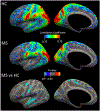
Comparison of sHIC between participants with MS and healthy controls. Average sHIC correlation maps are shown for the MS and HC groups. Significant differences in the MS vs HC groups were detected using a vertex-wise general linear model controlling for age and gender (cluster level p values < 0.05). For the p value color scale, blue/cold clusters represent cortical areas where sHIC was significantly reduced in the MS group. Gray color indicates that the p value was not significant. There were no areas displaying higher sHIC (red/hot clusters) in the MS group
Fig. 3
Fig. 3
Partial regression plot. This demonstrates the relationship between global sHIC and microstructural alterations in the posterior segment of the CC as measured by a restricted volume fraction (RVF) and b fractional anisotropy (FA), adjusting for age and gender. The partial correlation and adjusted p value is reported on each plot
Fig. 4
Fig. 4
Associations between sHIC and EDSS in participants with MS. Clusters were generated from a vertex-wise general linear model controlling for age and gender (cluster level p values < 0.05). The partial correlation values are displayed for the clusters that survived correction for multiple comparison. Blue/cold clusters represent cortical areas where there was a significant negative association between sHIC and EDSS. Gray color indicates that the p-value was not significant. There were no areas displaying a positive association between sHIC and EDSS
Fig. 5
Fig. 5
The interaction between posterior CC damage and posterior sHIC in predicting clinical disability. To provide a better understanding and visualization of the interaction effect, participants with MS were divided equally into two groups (low and high sHIC) based on the median group value for posterior sHIC. Regression lines are plotted for each group to show the trends for subjects with low and high posterior sHIC. For the group with low posterior sHIC (red) there was a positive association between posterior CC damage, measured by restricted volume fraction (RVF), and clinical disability and cognitive dysfunction as measured by (a) EDSS and (c) BVMT. This positive association was attenuated for the group with higher posterior sHIC (blue). This phenomenon was also observed for fractional anisotropy (FA) in the posterior CC and the clinical measures (b) EDSS and (d) BVMT. The interaction term beta and p-values from each regression model are reported on the corresponding plots

References

    1. Anticevic A, Dierker DL, Gillespie SK, Repovs G, Csernansky JG, Van Essen DC, Barch DM (2008) Comparing surface-based and volume-based analyses of functional neuroimaging data in patients with schizophrenia. Neuroimage 41(3):835–848. 10.1016/j.neuroimage.2008.02.052 - DOI - PMC - PubMed
    1. Barnard RO, Triggs M (1974) Corpus callosum in multiple sclerosis. J Neurol Neurosurg Psychiatry 37(11):1259–1264. 10.1136/jnnp.37.11.1259 - DOI - PMC - PubMed
    1. Behrens TE, Berg HJ, Jbabdi S, Rushworth MF, Woolrich MW (2007) Probabilistic diffusion tractography with multiple fibre orientations: What can we gain? Neuroimage 34(1):144–155. 10.1016/j.neuroimage.2006.09.018 - DOI - PMC - PubMed
    1. Benedict RH, Cookfair D, Gavett R, Gunther M, Munschauer F, Garg N, Weinstock-Guttman B (2006) Validity of the minimal assessment of cognitive function in multiple sclerosis (MACFIMS). J Int Neuropsychol Soc 12(4):549–558. 10.1017/s1355617706060723 - DOI - PubMed
    1. Bergendal G, Martola J, Stawiarz L, Kristoffersen-Wiberg M, Fredrikson S, Almkvist O (2013) Callosal atrophy in multiple sclerosis is related to cognitive speed. Acta Neurol Scand 127(4):281–289. 10.1111/ane.12006 - DOI - PubMed