

3D Bioprinting of Human Hollow Organs
- PMID: 35536418
- PMCID: PMC9088731
- DOI: 10.1208/s12249-022-02279-9
3D Bioprinting of Human Hollow Organs
Abstract
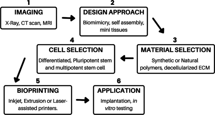
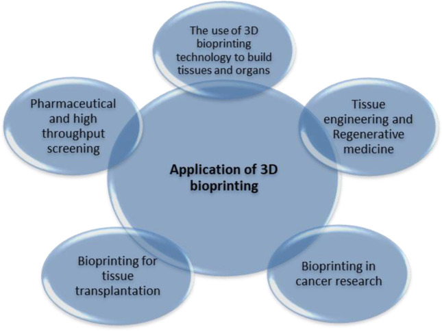
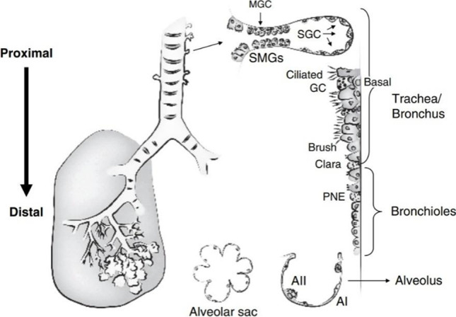
3D bioprinting is a rapidly evolving technique that has been found to have extensive applications in disease research, tissue engineering, and regenerative medicine. 3D bioprinting might be a solution to global organ shortages and the growing aversion to testing cell patterning for novel tissue fabrication and building superior disease models. It has the unrivaled capability of layer-by-layer deposition using different types of biomaterials, stem cells, and biomolecules with a perfectly regulated spatial distribution. The tissue regeneration of hollow organs has always been a challenge for medical science because of the complexities of their cell structures. In this mini review, we will address the status of the science behind tissue engineering and 3D bioprinting of epithelialized tubular hollow organs. This review will also cover the current challenges and prospects, as well as the application of these complicated 3D-printed organs.
Keywords: 3D bioprinting; bioinks; biomaterials; hollow organs.
© 2022. The Author(s), under exclusive licence to American Association of Pharmaceutical Scientists.
Conflict of interest statement
The authors declare no competing interests.
Figures
Similar articles
-
Recent Trends in Decellularized Extracellular Matrix Bioinks for 3D Printing: An Updated Review.Int J Mol Sci. 2019 Sep 18;20(18):4628. doi: 10.3390/ijms20184628. Int J Mol Sci. 2019. PMID: 31540457 Free PMC article. Review.
-
Advances in Regenerative Medicine and Biomaterials.Methods Mol Biol. 2023;2575:127-152. doi: 10.1007/978-1-0716-2716-7_7. Methods Mol Biol. 2023. PMID: 36301474
-
The fabrication of the chitosan-based bioink for in vitro tissue repair and regeneration: A review.Int J Biol Macromol. 2024 Feb;257(Pt 2):128504. doi: 10.1016/j.ijbiomac.2023.128504. Epub 2023 Nov 29. Int J Biol Macromol. 2024. PMID: 38040155 Review.
-
3D Tissue and Organ Printing-Hope and Reality.Adv Sci (Weinh). 2021 Mar 11;8(10):2003751. doi: 10.1002/advs.202003751. eCollection 2021 May. Adv Sci (Weinh). 2021. PMID: 34026444 Free PMC article. Review.
-
3D bioprinting of emulating homeostasis regulation for regenerative medicine applications.J Control Release. 2023 Jan;353:147-165. doi: 10.1016/j.jconrel.2022.11.035. Epub 2022 Nov 24. J Control Release. 2023. PMID: 36423869 Review.
Cited by
-
Advances in Regenerative Dentistry Approaches: An Update.Int Dent J. 2024 Feb;74(1):25-34. doi: 10.1016/j.identj.2023.07.008. Epub 2023 Aug 2. Int Dent J. 2024. PMID: 37541918 Free PMC article. Review.
-
AFM Analysis of a Three-Point Flexure Tested, 3D Printing Definitive Restoration Material for Dentistry.J Funct Biomater. 2023 Mar 10;14(3):152. doi: 10.3390/jfb14030152. J Funct Biomater. 2023. PMID: 36976076 Free PMC article.
-
A mathematical phase field model predicts superparamagnetic nanoparticle accelerated fusion of HeLa spheroids for field guided biofabrication.Sci Rep. 2025 Jun 5;15(1):19765. doi: 10.1038/s41598-025-04495-2. Sci Rep. 2025. PMID: 40473770 Free PMC article.
-
Human mini-blood-brain barrier models for biomedical neuroscience research: a review.Biomater Res. 2022 Dec 16;26(1):82. doi: 10.1186/s40824-022-00332-z. Biomater Res. 2022. PMID: 36527159 Free PMC article. Review.
-
Expanding Quality by Design Principles to Support 3D Printed Medical Device Development Following the Renewed Regulatory Framework in Europe.Biomedicines. 2022 Nov 16;10(11):2947. doi: 10.3390/biomedicines10112947. Biomedicines. 2022. PMID: 36428514 Free PMC article. Review.
References
-
- Kruth JP. Material increases manufacturing by rapid prototyping techniques. CIRP Ann. 1991;40(2):603–614. doi: 10.1016/S0007-8506(07)61136-6. - DOI
-
- Heller TB, Hill RM, Saggal AF. Inventors; Quadrax Corp, assignee. Apparatus for forming a solid three-dimensional article from a liquid medium. 1991. United States patent US 5,071,337.
-
- Nair LS, Laurencin CT. Polymers as biomaterials for tissue engineering and controlled drug delivery. Tissue Eng. 2005;I:47–90. - PubMed
-
- Hull CW. Inventor; UVP Inc, assignee. Apparatus for production of three-dimensional objects by stereolithography. Google Patents; 1986. US Patent 4,575,330.
Publication types
MeSH terms
LinkOut - more resources
Full Text Sources
