Review
. 2022 May 10;79(6):288.
doi: 10.1007/s00018-022-04290-6.

Intracellular mono-ADP-ribosyltransferases at the host-virus interphase

Affiliations
Review

Intracellular mono-ADP-ribosyltransferases at the host-virus interphase

Bernhard Lüscher et al. Cell Mol Life Sci. .

Abstract

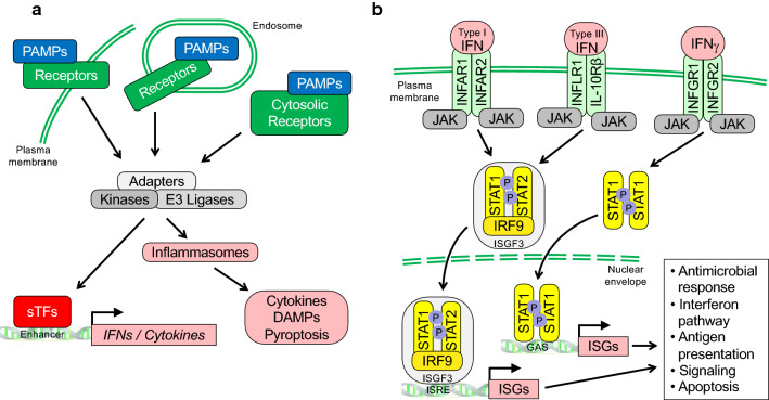
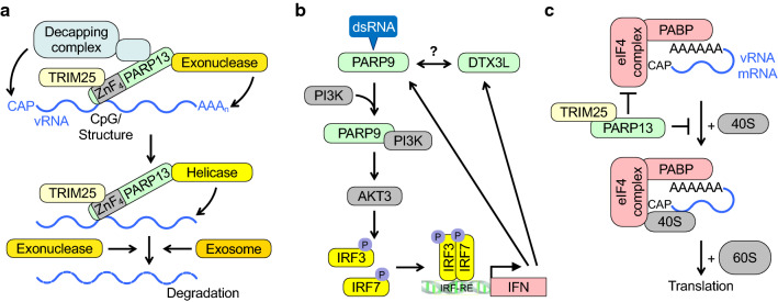
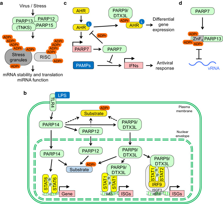
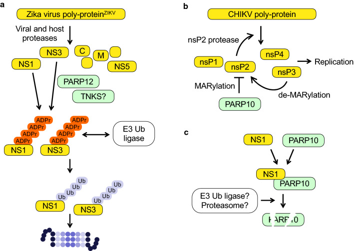
The innate immune system, the primary defense mechanism of higher organisms against pathogens including viruses, senses pathogen-associated molecular patterns (PAMPs). In response to PAMPs, interferons (IFNs) are produced, allowing the host to react swiftly to viral infection. In turn the expression of IFN-stimulated genes (ISGs) is induced. Their products disseminate the antiviral response. Among the ISGs conserved in many species are those encoding mono-ADP-ribosyltransferases (mono-ARTs). This prompts the question whether, and if so how, mono-ADP-ribosylation affects viral propagation. Emerging evidence demonstrates that some mono-ADP-ribosyltransferases function as PAMP receptors and modify both host and viral proteins relevant for viral replication. Support for mono-ADP-ribosylation in virus-host interaction stems from the findings that some viruses encode mono-ADP-ribosylhydrolases, which antagonize cellular mono-ARTs. We summarize and discuss the evidence linking mono-ADP-ribosylation and the enzymes relevant to catalyze this reversible modification with the innate immune response as part of the arms race between host and viruses.

Keywords: ADP-ribosylation; Alphavirus; Chikungunya virus; Coronavirus; Hydrolase; Interferon; MARylation; Macrodomain; PARP; Pattern recognition receptors; Signaling.


Conflict of interest statement

The authors declare that they have no competing interests.

Figures

Fig. 1
Fig. 1
Schematic summary of signaling processes in innate immunity. a Pathogen-associated molecular patterns (PAMPs) serve as markers recognized by pattern recognition receptors (PRRs) that allow cells to distinguish between self and non-self. PRRs include membrane bound Toll-like receptors (TLRs) and cytosolic nucleotide-binding oligomerization domain (NOD)-like receptors (NLRs), retinoic acid-inducible gene I (RIG-I)-like receptors (RLRs), AIM2-like receptors (ALRs), and the cyclic guanosine monophosphate–adenosine monophosphate synthase (cGAS). These receptors read different PAMPs, which include conserved microbial components such as glycolipids, peptidoglycans, lipopolysaccharides, and various nucleic acids such as dsRNA and dsDNA, and stimulate signaling complexes that typically involve adaptor proteins and enzymes with kinase and ubiquitin E3 ligase activity. Subsequently, these activate sequence specific transcription factors (sTFs) such as IFN regulatory factors (IRFs) and NF-kB proteins, and inflammasomes. The latter are multimolecular complexes controlling proteolytic enzymes such as caspase-1, which activate IL-1 family cytokines [260, 261]. As a consequence, IFNs, pro-inflammatory cytokines and alarmins or DAMPs (damage associated molecular patterns) are released and disseminate potentially hazardous pathogen encounters. b Different interferons (IFN) interact with distinct heterodimeric receptors as indicated. Upon cytokine binding, Janus family kinases (JAKs) are stimulated that phosphorylate transcription factors of the signal transducer and activator of transcription (STAT) family. Complexes of STAT1 and STAT2 with IRF9 form the trimeric transcription factor ISGF3, which binds to IFN-specific response elements (ISREs). Dimeric STAT1 complexes recognize IFNγ activation sites (GAS). Both ISREs and GAS elements are commonly found in IFN stimulated genes
Fig. 2
Fig. 2
PARPs regulate the stability and the translation of viral and cellular RNA. a The Zinc fingers (ZnF) of PARP13 sense CpG-rich RNA, possibly in the context of secondary structure elements. CpG-rich RNA is typically a hallmark of non-self RNA. This is enhanced by TRIM25. PARP13 also recruits exonucleases, decapping enzymes, helicases and the exosome, which together promote RNA degradation. b PARP9 senses viral dsRNA and promotes the phosphorylation and activation of IRF3 and 7 through PI3K and AKT3 and subsequently the activation of IFN genes. This activation is not dependent on signaling complexes used typically by PRRs. These signaling events may function as a feed-forward loop. Whether this activity of PARP9 requires DTX3L is unclear. c PARP13 inhibits translation of certain cellular and viral RNAs by interfering with the eukaryotic initiation factors and the binding of the 40S ribosomal subunit to mRNA. The specificity of these effects is not fully understood
Fig. 3
Fig. 3
Cellular processes controlled by PARP networks. a Different forms of stress, including viral infection, stimulate stress granule (SG) assembly. Several PARPs are associated with SGs. G3BP1 and 2 are ADP-ribosylated and these two proteins are necessary for SG formation and for replication of certain viruses (see text). Moreover, components of the RNA-induced silencing complex (RISC) are substrates of PARPs. Both SGs and RISC are controlling the stability and availability of mRNAs, processes targeted by viruses. b The interactions of the indicated IFN-inducible PARPs are summarized. The activation of TLR4 by LPS promotes the nuclear translocation of PARP14 as well as PARP12 and PARP9/DTX3L. A shift in substrate ADP-ribosylation from the cytosol to the nuclear compartment is proposed. Moreover, PARP14 and PARP9/DTX3L have cofactor function and influence ISG expression. Not shown is the suggested antagonism of PARP14 and PARP9 in regulating STAT1 (see text). c Ligands (L) such as xenobiotics and microbial metabolites activate AHR, which stimulates PARP7 expression. PARP7 in turn inhibits AHR function, at least in part through direct MARylation. MARylated AHR is read by PARP9 resulting in differential gene expression. PARP7 also modifies and inhibits TBK1 (not shown), a kinase activated by certain PAMPs, and thus prevents the expression of IFN genes. d PARP7 MARylates PARP13 at cysteines located in the ZnFs, which is suggested to interfere with the ability of PARP13 to sense CpG-rich vRNA
Fig. 4
Fig. 4
ADP-ribosylation of viral proteins and interaction with PARPs. a The non-structural proteins NS1 and NS2 of Zika virus are ADP-ribosylation substrates of PARP12 and potentially of a TNKS. The resulting PARylation promotes the recruitment of an E3 ubiquitin ligase. Subsequent poly-ubiquitination induces proteasomal degradation. At which point ADP-ribosylation is reversed has not been clarified. b The protease domain of the non-structural protein nsP2 of Chikungunya virus is MARylated by PARP10, which inhibits the proteolytic activity. As a result viral non-structural polyprotein processing and thus replication is inhibited. This is antagonized by the viral macrodomain of nsP3. c The non-structural protein NS1 of avian influenza virus interacts with PARP10 resulting in PARP10 degradation. Whether this involves poly-ubiquitination and proteasomal degradation has not been resolved

Similar articles

Cited by

References

    1. Chan YK, Gack MU. Viral evasion of intracellular DNA and RNA sensing. Nat Rev Microbiol. 2016;14(6):360–373. doi: 10.1038/nrmicro.2016.45. - DOI - PMC - PubMed
    1. O'Neill LA, Golenbock D, Bowie AG. The history of Toll-like receptors - redefining innate immunity. Nat Rev Immunol. 2013;13(6):453–460. doi: 10.1038/nri3446. - DOI - PubMed
    1. Chow KT, Gale M, Jr, Loo YM. RIG-I and other RNA sensors in antiviral immunity. Annu Rev Immunol. 2018;36:667–694. doi: 10.1146/annurev-immunol-042617-053309. - DOI - PubMed
    1. Zevini A, Olagnier D, Hiscott J. Crosstalk between cytoplasmic RIG-I and STING sensing pathways. Trends Immunol. 2017;38(3):194–205. doi: 10.1016/j.it.2016.12.004. - DOI - PMC - PubMed
    1. Ablasser A, Hur S. Regulation of cGAS- and RLR-mediated immunity to nucleic acids. Nat Immunol. 2020;21(1):17–29. doi: 10.1038/s41590-019-0556-1. - DOI - PubMed

Substances

LinkOut - more resources