

Dermoscopy of cutaneous adnexal tumours: a systematic review of the literature
- PMID: 35536546
- PMCID: PMC9545415
- DOI: 10.1111/jdv.18210
Dermoscopy of cutaneous adnexal tumours: a systematic review of the literature
Abstract
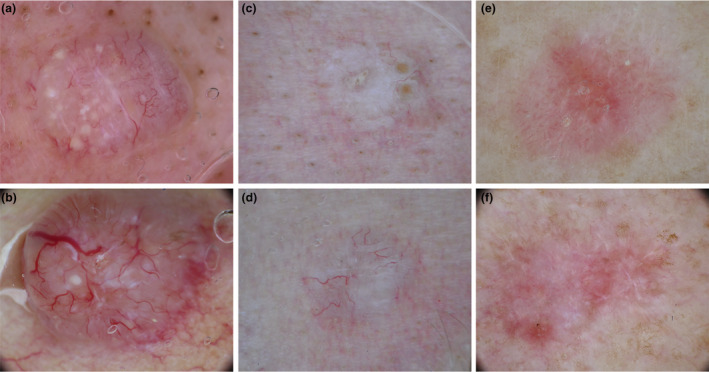
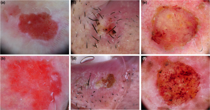
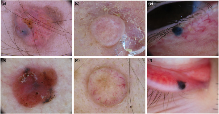
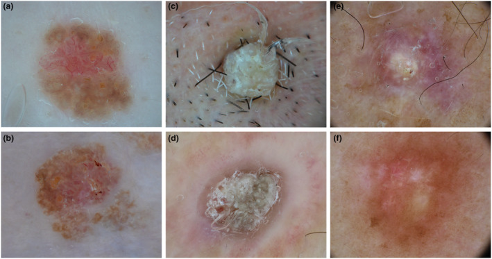
Cutaneous adnexal tumours (ATs) encompass a variegated group of hamartomas and benign or malignant tumours, originating from the hair follicle, sebaceous, eccrine or apocrine glands that may simulate other cutaneous neoplasms. This study aims to provide a comprehensive overview of the spectrum of clinical and dermoscopic features of ATs, to better define these lesions and assist in the differential diagnosis. We performed a two-step systematic search of the literature in PubMed, Embase and Cochrane Library databases from inception until 4 September 2020. In the first step, we aimed to define histological variants of ATs with descriptions of dermoscopic criteria. The second step included a search for the name of each previously identified AT variants in the same databases adding 'AND (epilum* or dermosc* or dermatosc*)'. All study types in English language reporting dermoscopic images of ATs were included. Collisions between ATs and other inflammatory or neoplastic skin lesions were excluded, with the exception of collisions with a sebaceous nevus. The protocol of this study was prospectively registered in PROSPERO (CRD42021244677). In total, 206 articles met our inclusion criteria, encompassing 372 ATs in 365 patients. Most ATs were apocrine-eccrine (n = 217, 58.3%, n = 173 benign) with a prevalence of poromas (n = 82), followed by follicular ATs (n = 88, 23.7%, n = 83 benign) and sebaceous ATs (n = 67, 18.0%, n = 49 benign). Most patients had a single AT lesion (320, 86.0%), while 42 (11.3%) had multiple ATs. A syndrome causing multiple ATs was identified in 15 patients. Histopathological analysis revealed 82% benign (n = 305) and 18.0% malignant (n = 67). ATs were classified according to their ability to mimic four groups of more common skin tumours: basal cell carcinoma, squamous cell carcinoma, melanocytic lesions and benign cutaneous lesions. Moreover, we have highlighted the ability of malignant variants of ATs to simulate benign skin lesions. This systematic review offers a comprehensive overview of the common clinical and dermoscopic features of follicular, sebaceous and apocrine-eccrine ATs and details possible differential dermoscopic features.
Keywords: adnexal skin tumours; apocrine; dermatoscopy; dermoscopy; eccrine; follicular; glandular; sebaceous.
© 2022 The Authors. Journal of the European Academy of Dermatology and Venereology published by John Wiley & Sons Ltd on behalf of European Academy of Dermatology and Venereology.
Figures
Similar articles
-
Dermoscopic and trichoscopic features of primary cutaneous lymphomas - systematic review.J Eur Acad Dermatol Venereol. 2021 Jul;35(7):1470-1484. doi: 10.1111/jdv.17219. Epub 2021 Apr 27. J Eur Acad Dermatol Venereol. 2021. PMID: 33710688
-
Interventions for infantile haemangiomas of the skin.Cochrane Database Syst Rev. 2018 Apr 18;4(4):CD006545. doi: 10.1002/14651858.CD006545.pub3. Cochrane Database Syst Rev. 2018. PMID: 29667726 Free PMC article.
-
Interventions for promoting habitual exercise in people living with and beyond cancer.Cochrane Database Syst Rev. 2018 Sep 19;9(9):CD010192. doi: 10.1002/14651858.CD010192.pub3. Cochrane Database Syst Rev. 2018. PMID: 30229557 Free PMC article.
-
Magnetic resonance perfusion for differentiating low-grade from high-grade gliomas at first presentation.Cochrane Database Syst Rev. 2018 Jan 22;1(1):CD011551. doi: 10.1002/14651858.CD011551.pub2. Cochrane Database Syst Rev. 2018. PMID: 29357120 Free PMC article.
-
Sertindole for schizophrenia.Cochrane Database Syst Rev. 2005 Jul 20;2005(3):CD001715. doi: 10.1002/14651858.CD001715.pub2. Cochrane Database Syst Rev. 2005. PMID: 16034864 Free PMC article.
Cited by
-
Extraocular cutaneous sebaceous carcinoma in a patient with Muir-Torre syndrome: special emphasis on histologic and dermoscopic features.Dermatol Reports. 2023 Sep 13;16(3):9832. doi: 10.4081/dr.2023.9832. eCollection 2024 Sep 2. Dermatol Reports. 2023. PMID: 39539987 Free PMC article.
-
Dermoscopy, Line-Field Confocal Optical Coherence Tomography, Reflectance Confocal Microscopy, and Ultra-High-Frequency Ultrasound: Clues for the Diagnosis of Hidrocystomas.Diagnostics (Basel). 2024 Nov 27;14(23):2671. doi: 10.3390/diagnostics14232671. Diagnostics (Basel). 2024. PMID: 39682579 Free PMC article.
-
Sebaceoma on the nose mimicking basal cell carcinoma: Pitfalls of dermoscopy and reflectance confocal microscopy.Skin Res Technol. 2022 Nov;28(6):886-888. doi: 10.1111/srt.13227. Epub 2022 Nov 2. Skin Res Technol. 2022. PMID: 36325590 Free PMC article.
-
MCATs: Case report of adnexal adenocarcinoma of not otherwise specified type.Clin Case Rep. 2023 Mar 15;11(3):e7097. doi: 10.1002/ccr3.7097. eCollection 2023 Mar. Clin Case Rep. 2023. PMID: 36937642 Free PMC article.
-
Clinical and Dermoscopic Characteristics of Cutaneous Chondroid Syringoma: A Systematic Review.Dermatol Pract Concept. 2024 Oct 30;14(4):e2024237. doi: 10.5826/dpc.1404a237. Dermatol Pract Concept. 2024. PMID: 39652927 Free PMC article. Review.
References
-
- Patterson JW. Tumors of cutaneous appendages. In Patterson JW, Hosler GA, Prenshaw KL, eds. Weedon's Skin Pathology, 5th edn. Elsevier, Edinburgh, Scotland, 2020: 951–1015 e25.
-
- Elder D, Massi D, Scolyer R, Willemze R. WHO Classification of Skin Tumours, Vol. 11, 4th edn. International Agency for Research on Cancer, Lyon, France, 2018.
-
- Zaballos P, Gómez‐Martín I, Martin JM, Bañuls J. Dermoscopy of adnexal tumors. Dermatolic Clin 2018; 36: 397–412. - PubMed
-
- Stroup DF, Berlin J, Morton S et al. Meta‐analysis of observational studies in epidemiology: a proposal for reporting. Meta‐analysis of observational studies in epidemiology (MOOSE) group. JAMA 2000; 283: 2008–2012. - PubMed
-
- Moher D, Liberati A, Tetzlaff J, Altman DG. Preferred reporting items for systematic reviews and meta‐analyses: the PRISMA statement. J Clin Epidemiol 2009; 62: 1006–1012. - PubMed
Publication types
MeSH terms
LinkOut - more resources
Full Text Sources
Medical
Miscellaneous
