. 2022 May;11(5):e12222.
doi: 10.1002/jev2.12222.

Extracellular vesicles enclosed-miR-421 suppresses air pollution (PM2.5 )-induced cardiac dysfunction via ACE2 signalling

Affiliations

Extracellular vesicles enclosed-miR-421 suppresses air pollution (PM2.5 )-induced cardiac dysfunction via ACE2 signalling

Hongyun Wang et al. J Extracell Vesicles. 2022 May.

Abstract

Air pollution, via ambient PM2.5, is a big threat to public health since it associates with increased hospitalisation, incidence rate and mortality of cardiopulmonary injury. However, the potential mediators of pulmonary injury in PM2.5 -induced cardiovascular disorder are not fully understood. To investigate a potential cross talk between lung and heart upon PM2.5 exposure, intratracheal instillation in vivo, organ culture ex vivo and human bronchial epithelial cells (Beas-2B) culture in vitro experiments were performed respectively. The exposed supernatants of Beas-2B were collected to treat primary neonatal rat cardiomyocytes (NRCMs). Upon intratracheal instillation, subacute PM2.5 exposure caused cardiac dysfunction, which was time-dependent secondary to lung injury in mice, thereby demonstrating a cross-talk between lungs and heart potentially mediated via small extracellular vesicles (sEV). We isolated sEV from PM2.5 -exposed mice serum and Beas-2B supernatants to analyse the change of sEV subpopulations in response to PM2.5 . Single particle interferometric reflectance imaging sensing analysis (SP-IRIS) demonstrated that PM2.5 increased CD63/CD81/CD9 positive particles. Our results indicated that respiratory system-derived sEV containing miR-421 contributed to cardiac dysfunction post-PM2.5 exposure. Inhibition of miR-421 by AAV9-miR421-sponge could significantly reverse PM2.5 -induced cardiac dysfunction in mice. We identified that cardiac angiotensin converting enzyme 2 (ACE2) was a downstream target of sEV-miR421, and induced myocardial cell apoptosis and cardiac dysfunction. In addition, we observed that GW4869 (an inhibitor of sEV release) or diminazene aceturate (DIZE, an activator of ACE2) treatment could attenuate PM2.5 -induced cardiac dysfunction in vivo. Taken together, our results suggest that PM2.5 exposure promotes sEV-linked miR421 release after lung injury and hereby contributes to PM2.5 -induced cardiac dysfunction via suppressing ACE2.

Keywords: ACE2; PM2.5; cardiac dysfunction; extracellular vesicles; miR-421.


Conflict of interest statement

The authors report no conflict of interest.

Figures
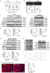
FIGURE 1
FIGURE 1
Cardiac dysfunction is secondary to lung injury and crosstalk occurred after PM2.5 exposure in vivo. (a) Schematic representation of experimental design in vivo, ex vivo and in vitro. To make subacute PM2.5 exposure model, mice received PM2.5 or PBS exposure via intratracheal instillation every day for 2 weeks (the first exposure day was labelled as day1 and the last exposure day was labelled as day14), followed by echocardiography measurement at day15, day21, day28 and day35, respectively. For ex vivo culture, the PBS or PM2.5 exposed mice lung and bronchi were dissected and the segments were cultured for 48 h. We then collected the ex vivo culture supernatants for use. For in vitro culture, the human bronchial epithelial cell line Beas‐2B exposed to PM2.5 for 48 h and collected the supernatants for centrifugation. Then the supernatants were collected for use. These supernatants were used to treat primary neonatal rat cardiomyocytes for 24 h. (b) Qualification of cardiac ejection fraction (EF) and fraction shorting (FS) at day35 in mice. n = 16. (c) Representative images obtained by western blot to show the protein expression of Bax, Bcl‐2, cleaved caspase 3 and caspase 3 in heart tissues lysates of mice. (d) Qualification of the ratio of Bax/Bcl‐2 and cleaved caspase 3/total caspase 3. n = 6. (e) Western blot images of Bax, Bcl‐2, cleaved caspase 3 and caspase 3 and (f) quantitation results of these proteins in Ex‐Ctr and Ex‐PM2.5 exposed NRCMs. n = 6. (g) Protein expression of Bax, Bcl‐2, cleaved caspase 3 and caspase 3 in SB‐Ctr or SB‐PM2.5 exposed NRCMs, n = 6. (h) TEM images and marker proteins of mice serum‐derived sEV. Scale bar = 100 nm (i) Concentration and size of serum‐derived sEV were tested by nanoparticle tracking analysis (Zetaview). (j) Representative images of TUNEL/α‐actinin/Hoechst staining of PBS‐sEV or PM2.5‐sEV exposed NRCMs. Scale bar = 50μm. (k) Quantitation of TUNEL positive NRCMs after PBS‐sEV or PM2.5‐sEV treatment. n = 5. Data were represented as mean ± SD. **p < 0.01 and ***p < 0.001, from comparing PBS‐ and PM2.5‐exposed groups, calculated by unpaired Student's t‐test (two‐sided). Ex‐Ctr : ex‐vivo culture supernatants of PBS‐exposed mice lung and bronchi. Ex‐PM2.5 : ex vivo culture supernatants of PM2.5‐exposed mice lung and bronchi. SB‐Ctr : Supernatants of PBS‐exposed Beas‐2B cells. SB‐PM2.5 : Supernatants of PM2.5‐exposed Beas‐2B cells. PBS‐sEV : Small EVs derived from PBS‐exposed mice serum. PM2.5‐sEV : Small EVs derived from PM2.5‐exposed mice serum
FIGURE 2
FIGURE 2
PM2.5 exposure alters extracellular particle subpopulations. (a) The morphology of SB‐ctr and SB‐PM2.5 derived EVs were displayed by TEM imaging. Scale bar = 50 nm. (b) Number of EV‐marker captured subpopulations (derived from PM2.5‐exposed Beas‐2B cell supernatant) were analysed by Single‐Particle Interferometric Reflectance Imaging Sensor (SP‐IRIS). (c) Protein marker such as CD63, CD9, Tsg101, Alix and negative marker (calnexin) were assessed by western blot. (d) Size distribution and (e) immune‐colocation of EVs were performed by SP‐IRIS. The chips were coated with antibodies against anti‐human CD81, CD63, CD9, respectively. Anti‐mouse IgG is the negative control. Fluorescent antibodies against CD81, CD63 and CD9 were used for colocation. Data were represented as mean ± SD. n = 3. ***p < 0.001 from comparing Ctr‐ and PM2.5‐exposed groups. SB‐Ctr‐EVs : EVs derived from Beas‐2B cell supernatants after exposure to PBS. SB ‐PM 2.5 ‐EVs : EVs derived from Beas‐2B cell supernatants after exposure to PM2.5 for 24 h. SB‐Ctr : Supernatants of PBS‐exposed Beas‐2B cells. SB‐PM 2.5 : Supernatants of PM2.5‐exposed Beas‐2B cells
FIGURE 3
FIGURE 3
GW4869 improves acute PM2.5‐induced cardiac dysfunction in vivo and in vitro. (a) Representative images of echocardiography measurement at day35 in PM2.5 or PM2.5+GW4869 treated mice. Quantitation of (b) Ejection fraction and fractional shortening (n = 16–18) and (c) TUNEL positive cardiomyocytes (CMs) (n = 5) in heart sections. Scale bar = 50μm. (d) Protein expression of Bax, Bcl‐2, cleaved caspase 3 and total caspase 3 by western blot and (e) relative quantitation results of the ratio of Bax/Bcl‐2, cleaved caspase 3/caspase 3 in mice heart tissues. n = 6. (f) Representative images of TUNEL/α‐actinin/Hoechst staining in NRCMs after exposure to Ex‐Ctr, Ex‐PM2.5 or Ex‐(PM2.5+GW4869) for 24 h and the quantitation results of TUNEL positive NRCMs. Scale bar = 50μm. n = 5. (g) Protein expression of Bax and Bcl‐2 after Ex‐Ctr, Ex‐PM2.5 and Ex‐(PM2.5+GW4869) treatment were assessed by western blot in NRCMs. n = 6. (h) TUNEL/α‐actinin/Hoechst staining in NRCMs after exposure to SB‐Ctr, SB‐GW4869, SB‐PM2.5 or SB‐(PM2.5+GW4869) for 24 h and TUNEL positive NRCMs were quantitated. n = 5. (i) Protein expression of Bax and Bcl‐2 were assessed by western blot in NRCMs after exposure to SB‐Ctr, SB‐GW4869, SB‐PM2.5 or SB‐(PM2.5+GW4869) for 24 h. n = 6. Data were represented as mean ± SD. *p < 0.05, **p < 0.01 and ***p < 0.001, respectively, from comparing control‐ and PM2.5‐exposed groups. p‐values were calculated by one way (b, f, g) or two‐way ANOVA (c, e, h and i) with Tukey correction for multiple comparisons. Ex‐Ctr : ex vivo culture supernatants of PBS‐exposed mice lung and bronchi. Ex‐PM 2.5 : ex vivo culture supernatants of PM2.5‐exposed mice lung and bronchi. Ex‐(GW+PM 2.5 ) : GW4869 were administrated to mice for 2 hours prior to exposure to PM2.5. Separating the lungs and bronchi from these mice at day15 for 48 h and then collected the supernatants. SB‐Ctr : Supernatants of PBS‐exposed Beas‐2B cells. SB‐PM 2.5 : Supernatants of PM2.5‐exposed Beas‐2B cells. SB‐GW4869 : Supernatants of GW4869 treated Beas‐2B cells. SB‐(GW+PM 2.5 ) : Beas‐2B cells were pretreated with GW4869 and then exposed to PM2.5 for 24 h. Collecting the supernatants for use
FIGURE 4
FIGURE 4
Respiratory system derived sEV packaging miR‐421 mediates cardiomyocytes apoptosis after PM2.5 exposure. Relative content of hsa‐miR‐421 in (a) Beas‐2B cells, (b) small EVs derived from the supernatants of Beas‐2B and (c) NRCMs. n = 5–6. 5S and U6 were used as internal control of cellular miR‐421 and sEV‐contained miR‐421, respectively. (d) Schematic representation of experimental design in vivo. To investigate the effect of miR‐421 in PM2.5‐induced cardiac dysfunction in vivo, AAV9‐miR421‐sponge and its control (AAV9‐Ctr) were utilised to inject into mice followed by PM2.5 exposure. Echocardiography (Echo) measurement were performed at day35. (e) The knockdown efficiency of mmu‐miR421‐3p in heart was examined by Realtime PCR and 5S was used as the control. n = 6. (f) The ejection fraction and fractional shortening of heart at day35 were shown. n = 7,7,12,12. (g) Quantitation of TUNEL positive cardiomyocytes were analysed in AAV9‐Ctr or AAV9‐miR421‐sponge injected mice heart with or without PM2.5 exposure. n = 6. (h) The expression of ACE2 and Bax/Bcl‐2 were examined in PM2.5‐exposed AAV9‐Ctr or AAV9‐miR421‐sponge injected mice heart. n = 6. (i) The NRCMs were exposed to SB‐NC‐Ctr, SB‐miR‐421inhibitor‐Ctr, SB‐(NC+PM2.5) or SB‐(miR‐421 inhibitor+PM2.5) for 24 h and then received TUNEL/α‐actinin/Hoechst staining (Scale bar = 50 μm) and TUNEL positive NRCMs were quantitated. n = 5. (j) The protein expression of Bax and Bcl‐2 were assessed by western blot and quantitated by Image J. n = 6. Data were represented as mean ± SD. *p < 0.05, **p < 0.01 and ***p < 0.001, respectively. p‐values were calculated by unpaired Student's t‐test (b, h), one‐way ANOVA (a, c), two‐way ANOVA with Tukey correction (e, f, g, i and j) for multiple comparisons. SB‐Ctr : Supernatants of PBS‐exposed Beas‐2B cells. SB‐PM 2.5 : Supernatants of PM2.5‐exposed Beas‐2B cells. SB‐NC‐Ctr : Beas‐2B cells were transfected with negative control siRNA followed by exposure to fresh PBS/DMEM and the supernatants were collected. SB‐miR421 inhibitor‐Ctr : Beas‐2B cells were transfected with miR‐421 inhibitor followed by exposure to fresh PBS/DMEM and the supernatants were collected. SB‐NC‐PM 2.5 : Beas‐2B cells were transfected with negative control siRNA followed by exposure to PM2.5 for 24 h and the supernatants were collected. SB‐miR421 inhibitor‐ PM 2.5 : Beas‐2B cells were transfected with miR‐421 inhibitor followed by exposure to PM2.5 for 24 h and the supernatants were collected
FIGURE 5
FIGURE 5
sEV‐miR421 targets cardiac ACE2 and mediates PM2.5‐induced cardiomyocytes apoptosis. (a) The bioinformatics prediction results for downstream of miR‐421 in human and mice through miRDB database. (b) To verify the binding site between miR‐421 and ACE2, we constructed luciferase reporter gene with higher luminescence in the ACE2 3′‐UTR and then cotransfected with miR‐421 mimic, which caused a significant decrease in the expression of luciferase. However, miR‐421 mimic has no effect on ACE2 3′‐UTR mutant luciferase expression. n = 6. (c) The protein expression of ACE2 in mice heart were assessed by western blot at day36. GW4869 were administrated to mice via intraperitoneal injection every other day. n = 6. The protein expression of ACE2 in NRCMs after exposure to (d) Ex‐Ctr, Ex‐PM2.5 or Ex‐(PM2.5+GW4869) as well as (e) SB‐Ctr, SB‐GW4869, SB‐PM2.5 or SB‐(PM2.5+GW4869). n = 6. (f) Relative ACE2 expression in NRCMs after exposure to SB‐NC‐Ctr, SB‐miR421 inhibitor‐Ctr, SB‐NC‐PM2.5 or SB‐miR421 inhibitor‐PM2.5. n = 6. (g) Functional reverse experiment of miR‐421 in vitro. NRCMs were transfected with si‐ACE2 or negative control and then exposed to SB‐PM2.5 or SB‐miR421 inhibitor‐PM2.5 for 24 h. Subsequently, NRCMs apoptosis was assessed by TUNEL staining combined with α‐actinin/Hoechst staining. n = 5. Data were represented as mean ± SD. *p < 0.05, **p < 0.01 and ***p < 0.001, respectively. p‐values were calculated by one‐way ANOVA with Tukey correction (d) or two‐way ANOVA with Tukey correction (b, c, e, f and g) for multiple comparisons. Ex‐Ctr: ex vivo culture supernatants of PBS‐exposed mice lung and bronchi. Ex‐PM2.5 : ex vivo culture supernatants of PM2.5‐exposed mice lung and bronchi. Ex‐(GW+PM2.5): GW4869 were administrated to mice and then exposed to PM2.5. Separating the lungs and bronchi from these mice at day15 for 48 h and then collected the supernatants. SB‐Ctr : Supernatants of PBS‐exposed Beas‐2B cells. SB‐PM 2.5 : Supernatants of PM2.5‐exposed Beas‐2B cells. SB‐GW4869 : Supernatants of GW4869 treated Beas‐2B cells. SB‐(GW+PM 2.5 ) : Beas‐2B cells were pretreated with GW4869 and then exposed to PM2.5 for 24 h. Collecting the supernatants and centrifugating removal of pallets for use. SB‐NC‐Ctr : Beas‐2B cells were transfected with negative control siRNA followed by exposure to fresh PBS/DMEM and the supernatants were collected and centrifugated. SB‐miR421 inhibitor‐Ctr : Beas‐2B cells were transfected with miR‐421 inhibitor followed by exposure to fresh PBS/DMEM and the supernatants were collected. SB‐NC‐PM 2.5 : Beas‐2B cells were transfected with negative control siRNA followed by exposure to PM2.5 for 24 h and the supernatants were collected. SB‐miR421 inhibitor‐PM 2.5 : Beas‐2B cells were transfected with miR‐421 inhibitor followed by exposure to PM2.5 for 24 h and the supernatants were collected. Si‐ACE2 : ACE2‐siRNA
FIGURE 6
FIGURE 6
Upregulation of ACE2 attenuates PM2.5‐induced cardiomyocytes apoptosis in vitro and in vivo. NRCMs were transfected with over expressed‐human ACE2 (OE‐hACE2) or scramble control (Scr) followed by exposure to SB‐Ctr or SB‐PM2.5 for 24 h respectively and then received TUNEL/α‐actinin/Hoechst staining. (a) Representative images of TUNEL staining and (b) quantitation of TUNEL positive NRCMs. Scale bar = 50 μm. n = 5. (c) NRCMs were transfected with OE‐hACE2 or scramble control followed by exposure to SB‐Ctr or SB‐PM2.5 for 24 h respectively and relative expression of Bax and Bcl‐2 were assessed by western blot. n = 6. Diminazene aceturate (DIZE) was used to reveal the effect of ACE2 activation on PM2.5‐induced cardiac dysfunction. Cardiac function was examined by echocardiography at day35. (d) Representative images of cardiac function were obtained by echocardiography, and cardiac ejection fraction and fraction shorting were quantified. n = 11. (e) The protein expression of ACE2, Bax, Bcl‐2, cleaved caspase 3 and caspase 3 in heart tissue lysates of mice. n = 6. Data were represented as mean ± SD. *p < 0.05, **p < 0.01 and ***p < 0.001. p‐values were calculated by two‐way ANOVA with Tukey correction for multiple comparisons

Similar articles

Cited by

References

    1. Abohashem, S. , Osborne, M. T. , Dar, T. , Naddaf, N. , Abbasi, T. , Ghoneem, A. , Radfar, A. , Patrich, T. , Oberfeld, B. , Tung, B. , Fayad, Z. A. , Rajagopalan, S. , & Tawakol, A. (2021). A leucopoietic‐arterial axis underlying the link between ambient air pollution and cardiovascular disease in humans. European Heart Journal, 42, 761–772. 10.1093/eurheartj/ehaa982 - DOI - PMC - PubMed
    1. Arab, T. , Mallick, E. R. , Huang, Y. , Dong, L. , Liao, Z. , Zhao, Z. , Gololobova, O. , Smith, B. , Haughey, N. J. , Pienta, K. J. , Slusher, B. S. , Tarwater, P. M. , Tosar, J. P. , Zivkovic, A. M. , Vreeland, W. N. , Paulaitis, M. E. , & Witwer, K. W. (2021). Characterization of extracellular vesicles and synthetic nanoparticles with four orthogonal single‐particle analysis platforms. Journal of Extracellular Vesicles, 10, e12079. 10.1002/jev2.12079 - DOI - PMC - PubMed
    1. Atkinson, R. W. , Kang, S. , Anderson, H. R. , Mills, I. C. , & Walton, H. A. (2014). Epidemiological time series studies of PM2.5 and daily mortality and hospital admissions: a systematic review and meta‐analysis. Thorax, 69, 660–665. 10.1136/thoraxjnl-2013-204492 - DOI - PMC - PubMed
    1. Bai, L. , Shin, S. , Burnett, R. T. , Kwong, J. C. , Hystad, P. , Van Donkelaar, A. , Goldberg, M. S. , Lavigne, E. , Copes, R. , Martin, R. V. , Kopp, A. , & Chen, H. (2019). Exposure to ambient air pollution and the incidence of congestive heart failure and acute myocardial infarction: A population‐based study of 5.1 million Canadian adults living in Ontario. Environment International, 132, 105004. 10.1016/j.envint.2019.105004 - DOI - PubMed
    1. Barnes, P. J. , Burney, P. G. J. , Silverman, E. K. , Celli, B. R. , Vestbo, J. , Wedzicha, J. A. , & Wouters, E. F. M. (2015). Chronic obstructive pulmonary disease. Nature Reviews Disease Primers, 1, 15076. 10.1038/nrdp.2015.76 - DOI - PubMed

Publication types