. 2022 Jun 22;7(12):e154722.
doi: 10.1172/jci.insight.154722.

Human CD4+CD8α+ Tregs induced by Faecalibacterium prausnitzii protect against intestinal inflammation

Affiliations

Human CD4+CD8α+ Tregs induced by Faecalibacterium prausnitzii protect against intestinal inflammation

Sothea Touch et al. JCI Insight. .

Abstract

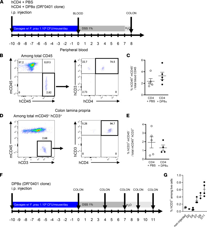
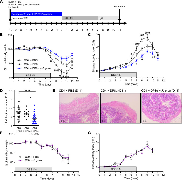
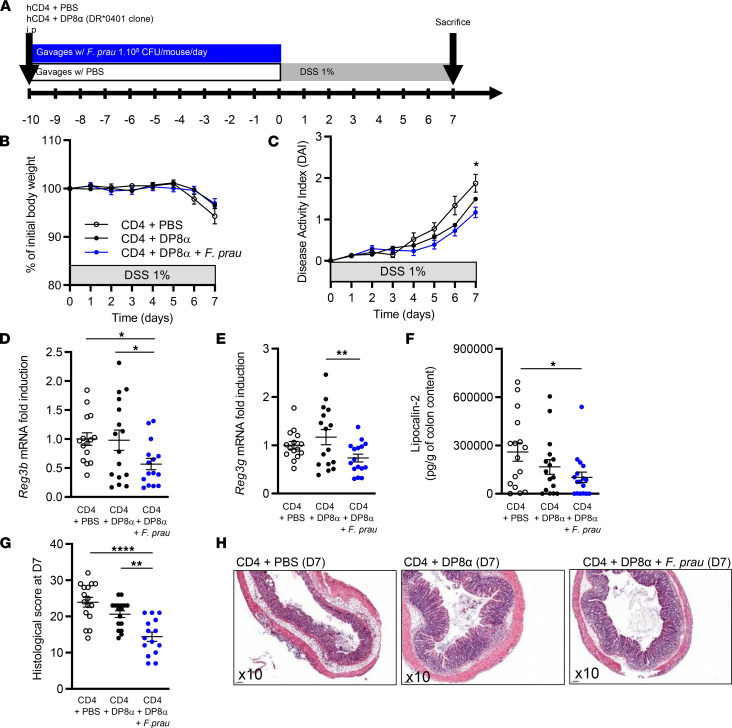
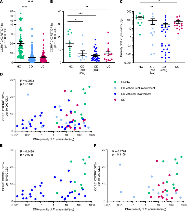
Abundance of Faecalibacterium prausnitzii, a dominant bacterium of the human microbiota that exhibits antiinflammatory effects, is decreased in patients with inflammatory bowel diseases (IBD). In humans, colonic lamina propria contains IL-10-secreting, Foxp3- Tregs characterized by a double expression of CD4 and CD8α (DP8α) and a specificity for F. prausnitzii. This Treg subset is decreased in IBD. The in vivo effect of DP8α cells has not been evaluated yet to our knowledge. Here, using a humanized model of a NSG immunodeficient mouse strain that expresses the HLA D-related allele HLA-DR*0401 but not murine class II (NSG-Ab° DR4) molecules, we demonstrated a protective effect of a HLA-DR*0401-restricted DP8α Treg clone combined with F. prausnitzii administration in a colitis model. In a cohort of patients with IBD, we showed an independent association between the frequency of circulating DP8α cells and disease activity. Finally, we pointed out a positive correlation between F. prausnitzii-specific DP8α Tregs and the amount of F. prausnitzii in fecal microbiota in healthy individuals and patients with ileal Crohn's disease.

Keywords: Gastroenterology; Inflammatory bowel disease; Mouse models; T cells.


Figures

Figure 1
Figure 1. Detection of human cells in whole blood and the colon lamina propria of NSG-Ab°DR4 mice.
(A) The presence of human cells after injection of CD4+ effector T cells in presence or absence of DP8α Tregs was assessed by flow cytometry in 2 independent experiments in the peripheral blood 10 days after injection, before the start of DSS treatment (day 0) and in the colon lamina propria 8 days after the start of DSS treatment (day 8). (B) After doublet discrimination and exclusion of debris, human CD3+ and human CD4+ cells were analyzed among human CD45+ (hCD45+) and murine CD45 (mCD45) cells. (C) Frequency of human CD45+ and mouse CD45 cells among total CD45+ cells in CD4+PBS (n = 5, empty dots) and CD4+DP8α (n = 5, black dots) groups. (D) Detection of human cells in the colon lamina propria was determined by the expression of human CD3 and the absence of expression of mouse CD45 among total mCD45+ and human CD3+ cells. (E) Frequency of human CD3+ and mouse CD45 cells among total colonic mouse CD45+ and human CD3+ cells in CD4+PBS (n = 5, empty dots) and CD4+DP8α (n = 4, black dots) groups. Original magnification, ×4. (F and G) DP8α clone was injected i.p. and analyzed by flow cytometry in the colon lamina propria of the mice after 10 days of daily gavage with F. prausnitzii followed by 0, 4, 7, 9, or 11 days of treatment with DSS. “Non-injected” indicates the control group without cells injected. Results are presented as the mean ± SEM.
Figure 2
Figure 2. Administration of DP8α Tregs and F. prausnitzii, but not that of DP8α or F. prausnitzii alone, protects NSG-Ab°DR4 mice from DSS-induced colitis.
(A) Experimental outline: NSG-Ab°DR4 female mice were injected i.p. with PBS or human peripheral CD4 effector T cells with either a human DRb1*0401-restricted DP8α Treg clone or PBS and received daily gavage with 1 × 10⁸ CFU of F. prausnitzii or 200 μL vehicle 1X PBS for 10 days before 1% DSS supplementation in drinking water for 7 days, followed by 4 days of regular drinking water. (B) Body weight and (C) disease activity index (DAI) were evaluated in the CD4+PBS (n = 44), CD4+DP8α (n = 20), and CD4+DP8α+F. prausnitzii (n = 45) groups in 5 independent experiments. (D and E) Histological score was assessed in the CD4+PBS (n = 30), CD4+DP8α (n = 9), and CD4+DP8α+F. prausnitzii (n = 29) groups in 3 independent experiments. (F) Body weight and (G) DAI were also evaluated in the CD4+PBS group (n = 10, open circles), as compared with the CD4+F. prausnitzii group (n = 11, purple circles) in another series of 2 independent experiments. Body weight and DAI are presented as the mean ± SEM. For comparison between multiple groups, 1-way ANOVA was performed, and P values of less than 0.05 were considered significant (*P < 0.05, **P < 0.01, ****P < 0.0001, §P < 0.05, §§P < 0.01, §§§P < 0.001, §§§§P < 0.0001). Only significant statistical results after adjustment for false discovery rate are shown (Benjamini-Hochberg, q < 0.1). The symbol † indicates the beginning of mortality.
Figure 3
Figure 3. Administration of DP8α Tregs and F. prausnitzii lowers inflammation induced by DSS-induced colitis in NSG-Ab°DR4 mice.
(A) Experimental outline: NSG-Ab°DR4 female mice were injected i.p. with 2 × 10⁶ human peripheral CD4 effector T cells alone or in combination with 2 × 10⁶ human DRb1*0401-restricted DP8α clones and received daily gavage with 200 μL 1X PBS or 1 × 10⁸ CFU of F. prausnitzii, respectively, for 10 days before 1% DSS supplementation in drinking water for 7 days. Mice were sacrificed at day 7. (B) Body weight and (C) disease activity index were assessed during the protocol in all groups of mice. (D and E) mRNA levels of Reg3b and Reg3g were analyzed by RT-qPCR in the proximal colon at day 7. (F) Lipocalin-2 secretion (pg/g of colon content) was measured by ELISA in the colon content at day 7. (G and H) Histological score was obtained from the colons of mice at day 7. Original magnification, ×10. Results are represented as the mean ± SEM. For comparison between multiple groups, 1-way ANOVA was performed, and P values of less than 0.05 were considered significant (*P < 0.05, **P < 0.01, ****P < 0.0001). Only significant statistical results after adjustment for false discovery rate are shown (Benjamini-Hochberg, q < 0.1). Each figure is representative of n = 3 independent experiments (CD4+PBS, n = 17; CD4+DP8α, n = 16; CD4+DP8α+F. prausnitzii, n = 17).
Figure 4
Figure 4. The frequency of circulating CCR6+CXCR6+ DP8α Tregs and the quantity of F. prausnitzii DNA in fecal microbiota are both decreased in patients with IBD and are positively correlated in patients with Crohn’s disease with ileal involvement.
(A) Frequency of circulating CCR6+CXCR6+ DP8α cells per 10,000 CD3+ T cells in the whole cohort of healthy controls (HC, n = 73), patients with Crohn’s disease (CD, n = 185), and patients with ulcerative colitis (UC, n = 65) or (B) according to localization (with or without ileal involvement) in a subcohort of HCs (n = 10) and patients with IBD (n = 10 CD without ileal involvement, n = 28 CD with ileal involvement, and n = 14 patients with UC). (C) Quantity of F. prausnitzii DNA in stool samples in HCs and patients with IBD. (D–F) Spearman’s correlation of the frequency of circulating CCR6+CXCR6+ DP8α cells per 10,000 CD3+ T cells and the quantity of F. prausnitzii DNA in fecal microbiota in the different groups. Results in A–C are represented as the mean ± SEM, For comparison between multiple groups, 1-way ANOVA was performed, and P values of less than 0.05 were considered significant (*P < 0.05, **P < 0.01, ***P < 0.001, ****P < 0.0001). Only significant statistical results after adjustment for false discovery rate are shown (Benjamini-Hochberg, q < 0.1).

References

    1. Goethel A, et al. The interplay between microbes and the immune response in inflammatory bowel disease. J Physiol. 2018;596(17):3869–3882. doi: 10.1113/JP275396. - DOI - PMC - PubMed
    1. Jostins L, et al. Host-microbe interactions have shaped the genetic architecture of inflammatory bowel disease. Nature. 2012;491(7422):119–124. doi: 10.1038/nature11582. - DOI - PMC - PubMed
    1. Khor B, et al. Genetics and pathogenesis of inflammatory bowel disease. Nature. 2011;474(7351):307–317. doi: 10.1038/nature10209. - DOI - PMC - PubMed
    1. Lamas B, et al. CARD9 impacts colitis by altering gut microbiota metabolism of tryptophan into aryl hydrocarbon receptor ligands. Nat Med. 2016;22(6):598–605. doi: 10.1038/nm.4102. - DOI - PMC - PubMed
    1. Liu JZ, et al. Association analyses identify 38 susceptibility loci for inflammatory bowel disease and highlight shared genetic risk across populations. Nat Genet. 2015;47(9):979–986. doi: 10.1038/ng.3359. - DOI - PMC - PubMed