. 2022 Jul 11;45(7):zsac110.
doi: 10.1093/sleep/zsac110.

Striatal mechanism of the restless legs syndrome

Affiliations

Striatal mechanism of the restless legs syndrome

Yuan-Yang Lai et al. Sleep. .

Abstract

Study objectives: Brain iron deficiency has been reported to be associated with the restless legs syndrome (RLS). However, 30%-50% of RLS patients do not respond to iron therapy, indicating that mechanisms other than brain iron deficiency may also participate in this disease. The striatum is known to be involved in the modulation of motor activity. We speculated that dysfunction of the striatum may induce RLS.

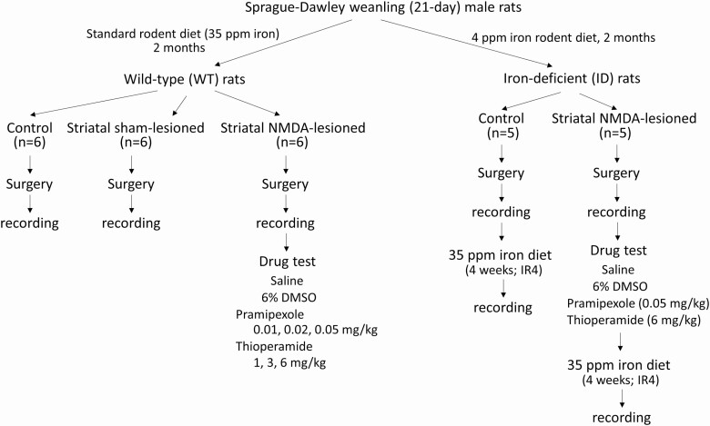
Methods: Two groups, wild-type (WT) and iron-deficient (ID) rats were used. Each group was divided into two subgroups, control and N-methyl-d-aspartate striatal-lesioned. After baseline recording, striatal-lesioned wild-type (WT-STL) and striatal-lesioned iron-deficient (ID-STL) rats were given pramipexole and thioperamide injections. Iron-deficient and ID-STL rats were then given a standard rodent diet for 4 weeks, and their sleep and motor activity were recorded.

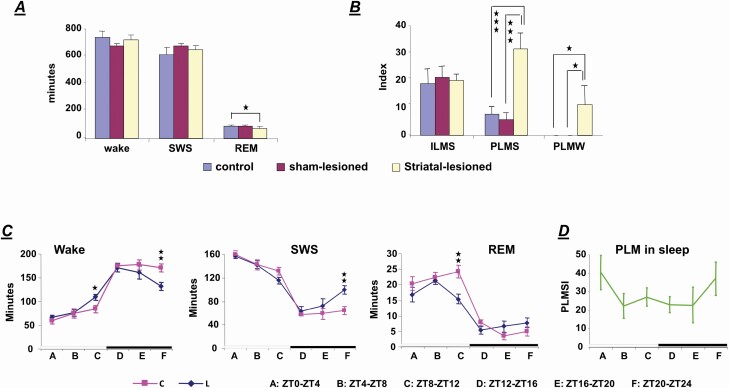
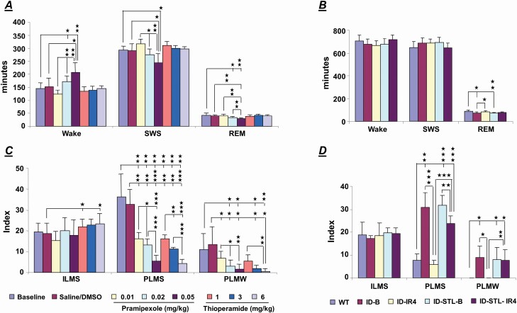
Results: WT-STL rats showed periodic leg movements (PLM) in wake, an increase in PLM in slow wave sleep (SWS), a decrease in rapid-eye-movement sleep, and a decrease in the daily average duration of episodes in SWS. The sleep-wake pattern and motor activity did not differ between ID and ID-STL rats. Thioperamide or pramipexole injection decreased PLM in sleep and in wake in WT-STL rats and ID-STL rats. Unlike ID rats, whose motor hyperactivity can be reversed by iron replacement, PLM in wake and in sleep in ID-STL rats were not fully corrected by iron treatment.

Conclusions: Lesions of the striatum generate RLS-like activity in rats. Dysfunction of the striatum may be responsible for failure to respond to iron treatment in some human RLS patients.

Keywords: iron deficient; iron therapy; neurotoxic lesions; periodic leg movement; pramipexole; thioperamide.


Figures

Figure 1.
Figure 1.
Diagram showing the experimental design. Two groups, wild-type (WT) and iron-deficient (ID), of male rats were used for the study. IR4: iron replacement for 4 weeks. See details in the Method section.
Figure 2.
Figure 2.
(A) Photomicrographs showing the lesion site in the striatum. (B) Reconstructed histology showing the NMDA-lesioned sites in the striatum. (C) Higher magnification photomicrograph, taken of the square area shown in (A), showing the lesion area circled with black dots. The arrow shown in (C) indicates the injection needle tract. Brain tissue was stained with cresyl violet. (D) and (E) example of periodic leg movements (PLM) in quiet wake (QW; D) and in sleep (E) observed in the striatal-lesioned wild-type rats. (F) example of PLM recorded in wild-type rat. EEG: electroencephalogram, L-EMG: leg electromyogram, OX: optic tract, PO: preoptic area, SPN: septal nucleus, ST: striatum. Calibration (C) 90 μL.
Figure 3.
Figure 3.
Effect of striatal lesions on sleep–wake pattern and motor activity in wild-type (WT) rats. (A) A decrease in rapid-eye-movement (REM) sleep with no change in wake and slow wave sleep (SWS) was found in the striatal-lesioned wild-type (WT-STL) rats in 24 hour recordings. (B) Striatal lesions in the WT rat produced an increase in periodic leg movements (PLM) in sleep (PLMS), as well as generating PLM in quiet wake (PLMW) in 24 hour recordings. In contrast, isolated leg movements in sleep (ILMS) were not changed in the WT-STL rats. Indexes of PLMS and PLMW were calculated as the total number of PLM in 24 hours in sleep and wake divided by the total time in sleep and wake in 24 hours. Index of ILMS was calculated as the total number of isolated motor events in 24 hours divided by the total time in sleep in 24 hours. (C) Distribution of wake, SWS and REM sleep time in 4-hour epoch over 24-hour recording in the WT control (C) and WT striatal-lesioned (L) rats. (D) Distribution of PLMS index (PLMSI) over 24-hour recording in the striatal-lesioned wild-type rats. Periodic leg movements in sleep were maximal at Zeitgeber time (ZT) 0–4 and ZT19–ZT23. The white and black bars shown on the bottom (C and D) represent the light and dark phases, respectively. t-Test, df = 10, *p < .05, **p < .01, ***p < .001.
Figure 4.
Figure 4.
Effect of pramipexole and thioperamide on sleep–wake pattern (A) and motor activity (C) in the striatal-lesioned wild-type (WT-STL) rats. Data were taken 8 hours after test drug injection. High doses of pramipexole (0.02 mg/kg and 0.05 mg/kg) injection into the WT-STL rats produced an increase in wake and a decrease in slow wave sleep (SWS) and rapid eye movement (REM) sleep (A). Thioperamide injected into the WT-STL rats had no effect on the sleep–wake pattern (A). Motor hyperactivity, PLM in sleep (PLMS) and in quiet wake (PLMW), was suppressed by pramipexole or thioperamide injection into the WT-STL rats (C). Effect of striatal lesions and iron replacement (IR) in the iron-deficient (ID) rat on sleep–wake pattern and motor activity in 24 hour recording. (B) The total time in wake and SWS in 24 hour recording did not differ between wild-type (WT), ID, and striatal-lesioned ID (ID-STL), as well as ID (ID-IR4) and ID-STL (ID-STL-IR4) rats fed standard rodent diet for 4 weeks. The total amount of REM sleep time in ID and ID-STL rats was lower than that in WT rats; however, the lower amount of REM sleep time in ID and ID-STL rats was corrected by iron replacement (IR). (D) Iron replacement decreased PLMS in both ID and ID-STL rats. Iron replacement decreased PLMW in the ID rats, but not in the ID-STL rats. Furthermore, iron replacement fully corrected PLMS and PLMW in the ID rats, but not the ID-STL rats. Indexes of PLMS and PLMW were calculated as the total number of PLM in 8 (B) and 24 (D) hours in sleep and wake divided by the total time in sleep and wake in 8 (B) and 24 (D) hours, respectively. Index of ILMS was calculated as the total number of isolated motor events in 8 (B) and 24 (D) hours divided by the total time in sleep in 8 (B) and 24 (D) hours, respectively. ID-B: baseline of iron-deficient rat, ID-STL-B: baseline of striatal-lesioned iron-deficient rat, W: wake. t-Test, * p < .05, ** p < .01, *** p < .001. Number of animals: wild-type: 6, iron-deficient and striatal-lesioned iron-deficient: 5 each.

Similar articles

Cited by

References

    1. Joo EY, et al. . Reduced cerebral blood perfusion of putamen and insular cortex in patients with idiopathic restless legs syndrome. J Korea Sleep Res Soc. 2012;9(1):10–14. doi:10.13078/jksrs.12003. - DOI
    1. Earley CJ, et al. . Increased synaptic dopamine in the putamen in restless legs syndrome. Sleep 2013;36(1):51–57. doi:10.5665/sleep.2300. - DOI - PMC - PubMed
    1. Michaud M, et al. . SPECT imaging of striatal pre- and postsynaptic dopaminergic status in restless legs syndrome with periodic leg movements in sleep. J Neurol. 2002;249(2):164–170. doi:10.1007/pl00007859. - DOI - PubMed
    1. Connor JR, et al. . Altered dopaminergic profile in the putamen and substantia nigra in restless leg syndrome. Brain 2009;132(9):2403–2412. doi:10.1093/brain/awp125. - DOI - PMC - PubMed
    1. Lai YY, et al. . Substantia nigra pars reticulata-mediated sleep and motor activity regulation. Sleep 2021;44(1):1–8. doi:10.1093/sleep/zsaa151. - DOI - PMC - PubMed

Publication types