

Modulation of dorsolateral prefrontal cortex functional connectivity after intermittent theta-burst stimulation in depression: Combining findings from fNIRS and fMRI
- PMID: 35537216
- PMCID: PMC9118162
- DOI: 10.1016/j.nicl.2022.103028
Modulation of dorsolateral prefrontal cortex functional connectivity after intermittent theta-burst stimulation in depression: Combining findings from fNIRS and fMRI
Abstract
Background: Resting-state functional magnetic resonance imaging (fMRI) can assess modulation of functional connectivity networks following repetitive transcranial magnetic stimulation (rTMS) in the treatment of depression. Functional near-infrared spectroscopy (fNIRS) is well suited for the concurrent application during rTMS treatment sessions to capture immediate blood oxygenation (oxy-Hb) effects, however limited in spatial resolution.
Objective: To understand the network effects behind such a prefrontal fNIRS response during rTMS, and to test whether the fNIRS signal may be predictive of treatment response, we linked data from fNIRS and fMRI within a clinical intervention study.
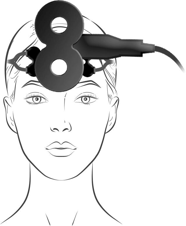
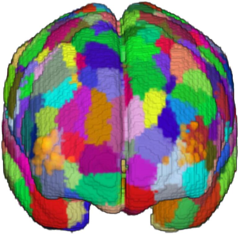
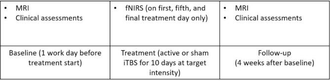
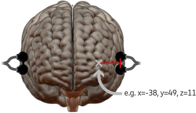
Methods: 42 patients with ongoing depression were recruited and randomized to receive active or sham intermittent theta-burst stimulation (iTBS) over the dorsomedial prefrontal cortex (dmPFC) twice daily for ten days at target intensity. Oxy-Hb was recorded with fNIRS during the first, fifth, and final day of iTBS, with the probe holders located laterally to the TMS coil over regions corresponding to the left and right dorsolateral prefrontal cortex (dlPFC). Resting-state fMRI scanning was performed before and after the whole iTBS treatment course. Functional connectivity analyses were then performed using dlPFC seeds from parcels of a brain atlas showing most overlap with the fNIRS probe locations during treatment.
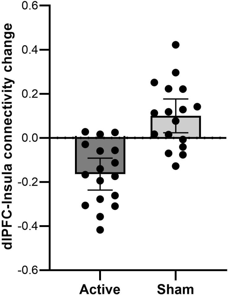
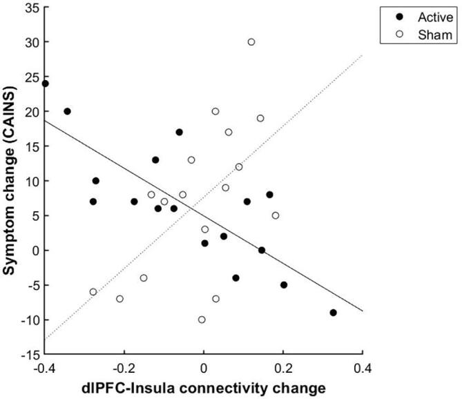
Results: After active iTBS, left dlPFC-connectivity to the right insula/operculum was reduced compared to sham. The left insula showed a connectivity reduction to the left dlPFC that correlated with an improvement in symptoms. In addition, the posterior parietal cortex showed a connectivity reduction to the left dlPFC that correlated with the fNIRS signal following active iTBS. Finally, the fNIRS oxy-Hb signal from the left dlPFC-seed during the first treatment day was predictive of dlPFC-connectivity change to precentral and temporal cortex regions.
Conclusion: By linking findings from these two different methods, this study suggests that changes within both the salience network and the central executive network affect the fNIRS response to iTBS.
Keywords: Central executive network; Depressive disorder; Repetitive transcranial magnetic stimulation (rTMS); Resting-state; Salience network.
Copyright © 2022 The Authors. Published by Elsevier Inc. All rights reserved.
Conflict of interest statement
The authors declare that they have no known competing financial interests or personal relationships that could have appeared to influence the work reported in this paper.
Figures
References
-
- Blumberger D.M., Vila-Rodriguez F., Thorpe K.E., Feffer K., Noda Y., Giacobbe P., Knyahnytska Y., Kennedy S.H., Lam R.W., Daskalakis Z.J., Downar J. Effectiveness of theta burst versus high-frequency repetitive transcranial magnetic stimulation in patients with depression (THREE-D): a randomised non-inferiority trial. The Lancet. 2018;391(10131):1683–1692. doi: 10.1016/S0140-6736(18)30295-2. - DOI - PubMed
-
- Bodén R., Bengtsson J., Thörnblom E., Struckmann W., Persson J. Dorsomedial prefrontal theta burst stimulation to treat anhedonia, avolition, and blunted affect in schizophrenia or depression – a randomized controlled trial. J. Affect. Disord. 2021;290(May):308–315. doi: 10.1016/j.jad.2021.04.053. - DOI - PubMed
Publication types
MeSH terms
LinkOut - more resources
Full Text Sources
Medical
