Case Reports
. 2022 May 10;15(5):e248543.
doi: 10.1136/bcr-2021-248543.

Chronic lymphoedema: a nidus for squamous cell carcinoma

Affiliations
Case Reports

Chronic lymphoedema: a nidus for squamous cell carcinoma

Shrea Gulati et al. BMJ Case Rep. .

Abstract

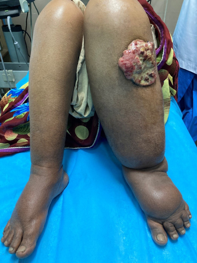
Lymphoedema is a chronic debilitating condition characterised by diffuse swelling caused by lymphatic obstruction. The secondary form of lymphoedema is more common than the primary form. Untreated filariasis remains an important cause of lymphoedema in developing countries. The most common complication of chronic lymphoedema is cellulitis. It is also a risk factor for the development of neoplasms such as lymphangiosarcoma, squamous cell carcinoma, melanoma, lymphoma and malignant fibrous histiocytoma. We report a case of a woman in her 60s who developed squamous cell carcinoma in the background of chronic lymphoedema.

Keywords: Dermatological; Skin cancer.


Conflict of interest statement

Competing interests: None declared.

Figures

Figure 1
Figure 1
Clinical image of a woman in her 60s with disproportionate lymphoedema of the left lower limb who developed a cauliflower-like, ulceroproliferative growth on the left shin, raising suspicion of a malignant ulcer.
Figure 2
Figure 2
Contrast CT scan (A) axial and (B) oblique multiplanar reconstruction images show an exophytic ulceroproliferative growth arising from the skin along the anteromedial aspect of the left shin, arising in the background of a swollen limb with extensive fat stranding (lymphoedema). There is no invasion of the muscles or bone by the growth. (C) Axial image at the level of inguinal ligament shows no significant lymphadenopathy. (D) Contrast-enhanced CT of the chest did not reveal any metastatic lung nodules.
Figure 3
Figure 3
Postoperative image shows healed surgical wound on the thigh with up to 30% graft uptake. The rest of the region shows epithelialisation.

Similar articles

Cited by

References

    1. Vignes S. [Lymphedema: From diagnosis to treatment]. Rev Med Interne 2017;38:97–105. 10.1016/j.revmed.2016.07.005 - DOI - PubMed
    1. Depairon M, Lessert C, Tomson D, et al. . [Primary lymphedema]. Rev Med Suisse 2017;13:2124–8. - PubMed
    1. Echenique-Elizondo M, Elorza J. Squamous-Cell carcinoma on long-lasting lymphoedema. Lancet Oncol 2002;3:319. 10.1016/S1470-2045(02)00734-9 - DOI - PubMed
    1. Parthiban R, Kaler AK, Shariff S, et al. . Squamous cell carcinoma arising from congenital lymphedema. SAGE Open Med Case Rep 2013;1:2050313X13496507:2050313X1349650. 10.1177/2050313X13496507 - DOI - PMC - PubMed
    1. Lister RK, Black MM, Calonje E, et al. . Squamous cell carcinoma arising in chronic lymphoedema. Br J Dermatol 1997;136:384–7. 10.1046/j.1365-2133.1997.d01-1206.x - DOI - PubMed

Publication types