. 2022 Aug;33(8):1487-1500.
doi: 10.1681/ASN.2022010048. Epub 2022 May 10.

Myeloid CCR2 Promotes Atherosclerosis after AKI

Affiliations

Myeloid CCR2 Promotes Atherosclerosis after AKI

Anne M Hüsing et al. J Am Soc Nephrol. 2022 Aug.

Abstract

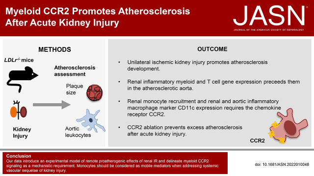
Background: The risk of cardiovascular events rises after AKI. Leukocytes promote atherosclerotic plaque growth and instability. We established a model of enhanced remote atherosclerosis after renal ischemia-reperfusion (IR) injury and investigated the underlying inflammatory mechanisms.

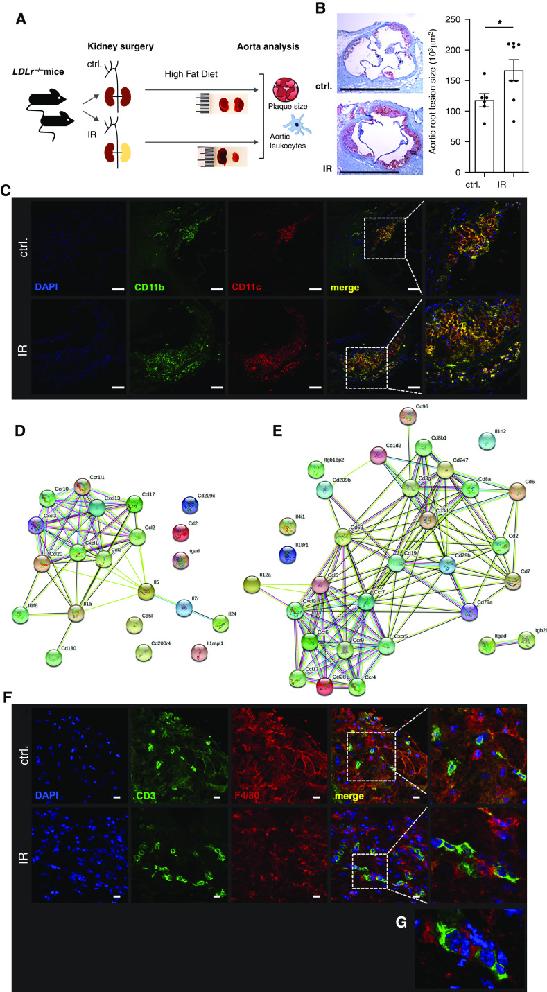
Methods: Atherosclerotic lesions and inflammation were investigated in native and bone marrow-transplanted LDL receptor-deficient (LDLr-/- ) mice after unilateral renal IR injury using histology, flow cytometry, and gene expression analysis.

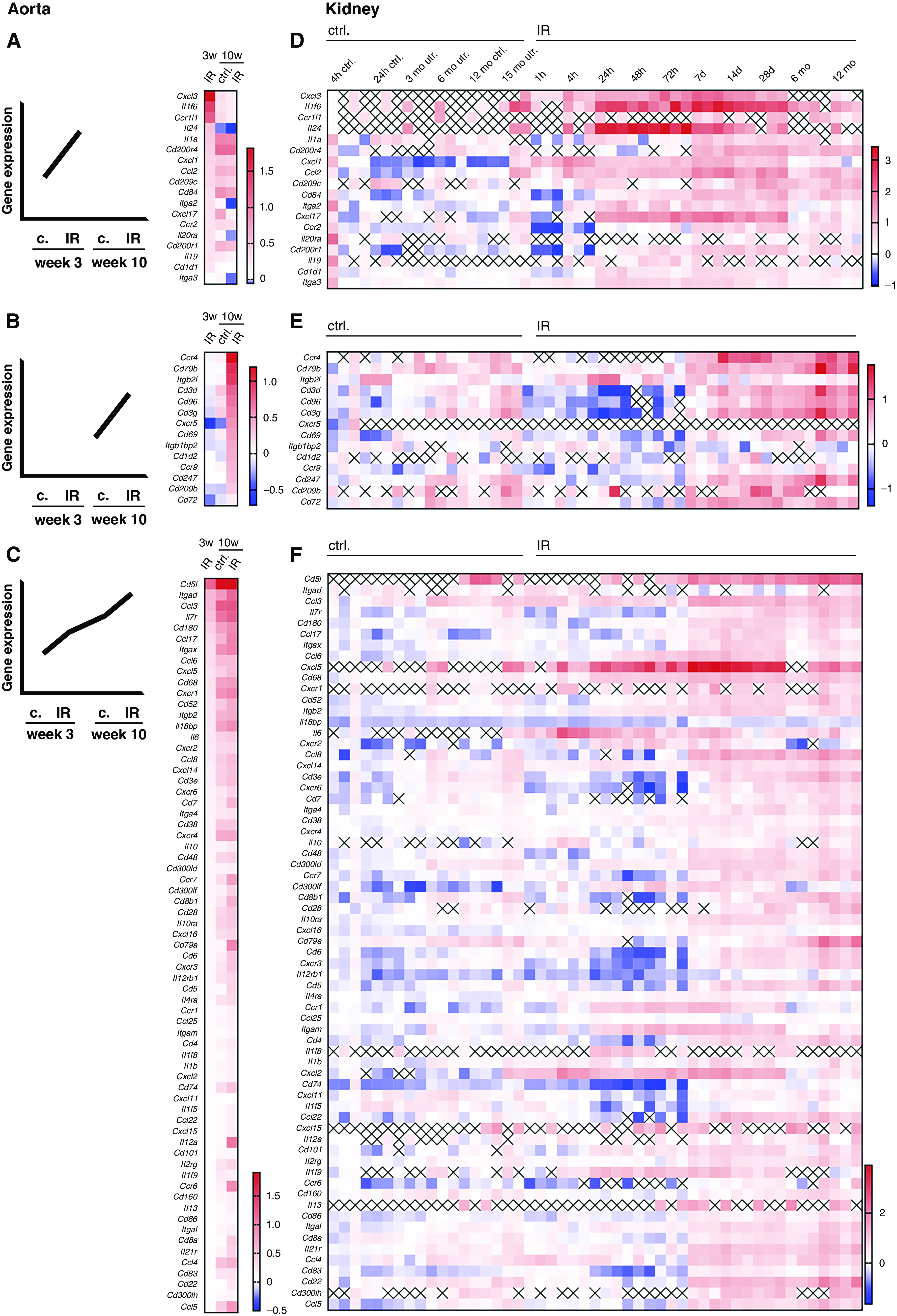
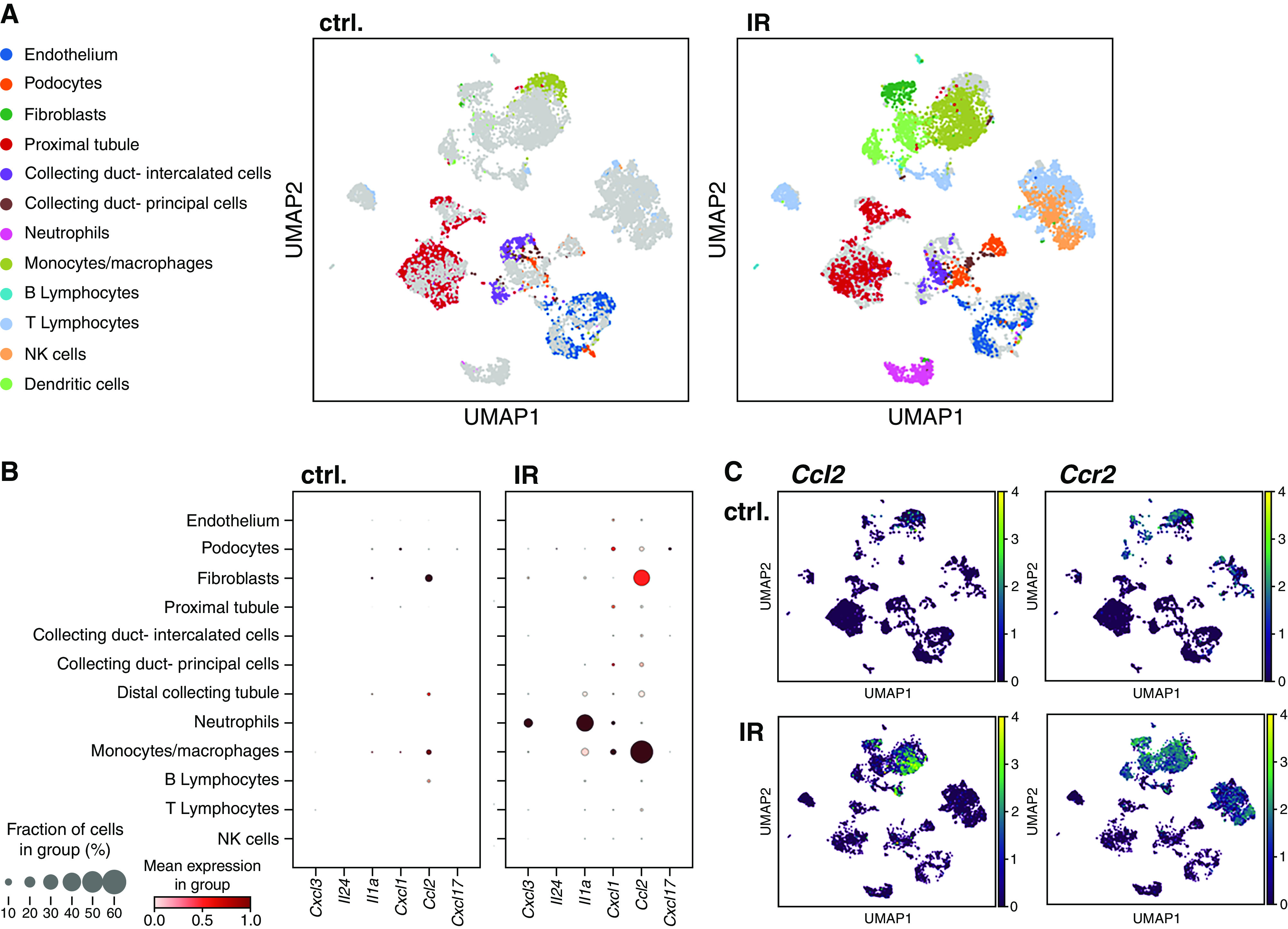
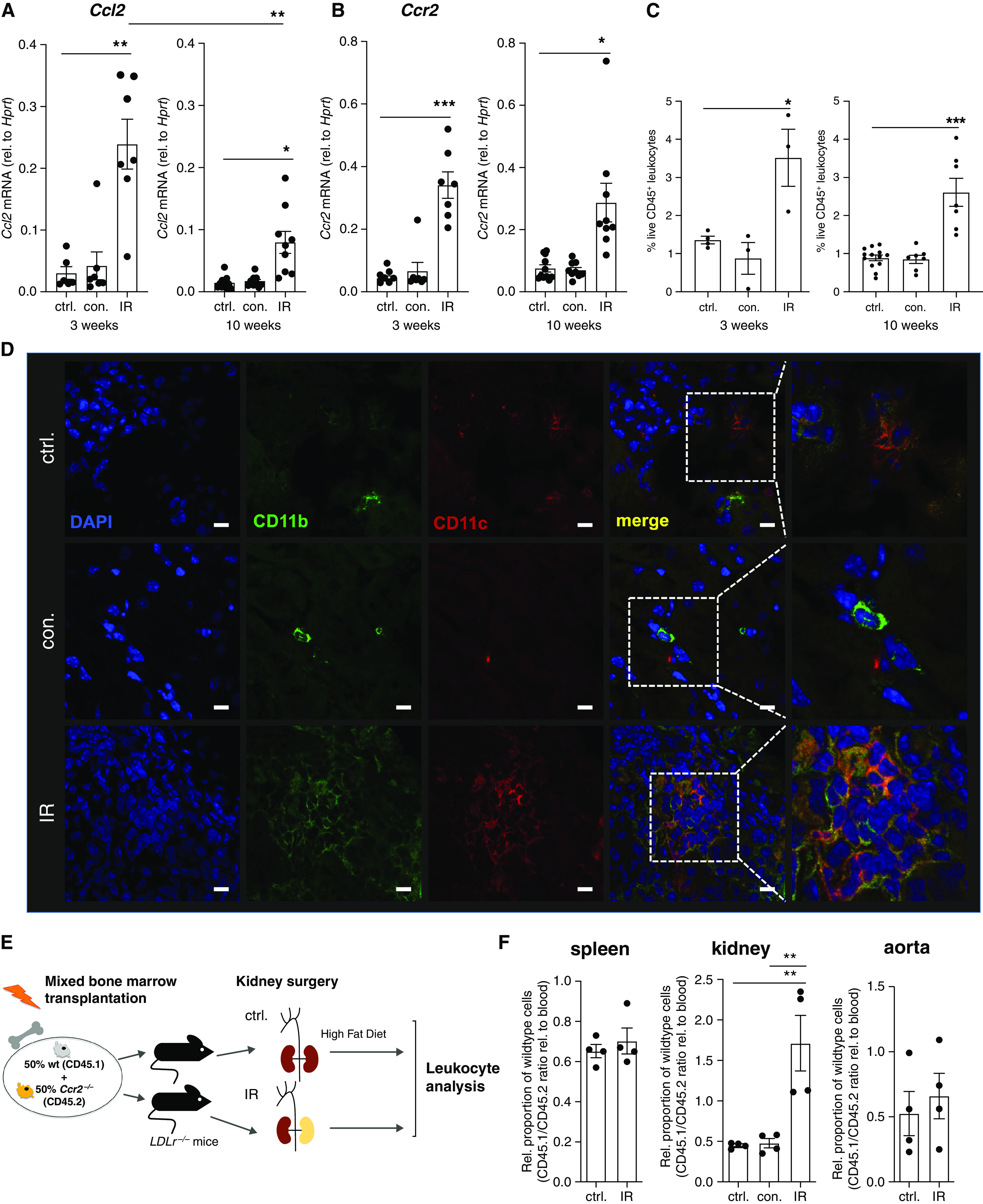
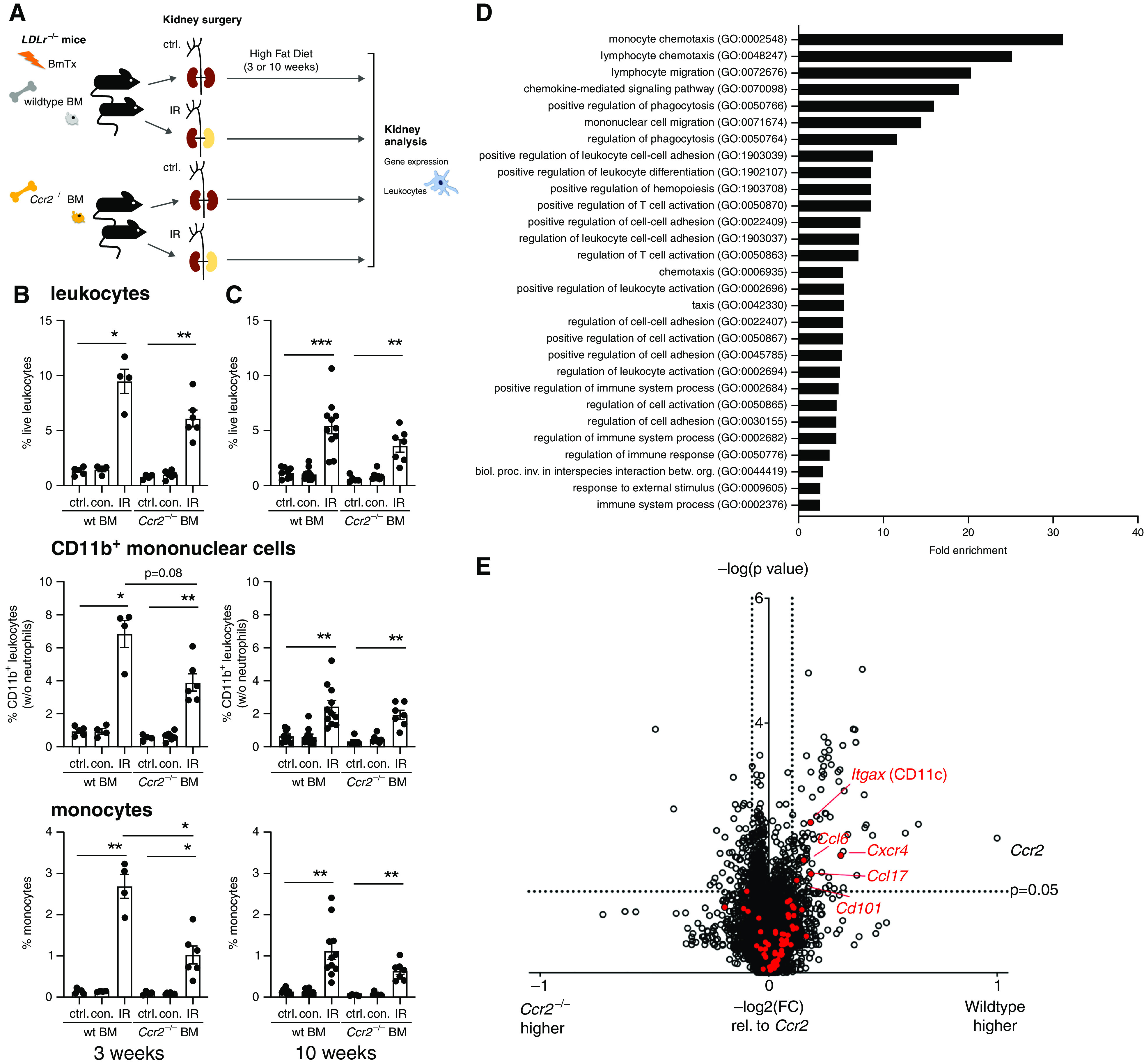
Results: Aortic root atherosclerotic lesions were significantly larger after renal IR injury than in controls. A gene expression screen revealed enrichment for chemokines and their cognate receptors in aortas of IR-injured mice in early atherosclerosis, and of T cell-associated genes in advanced disease. Confocal microscopy revealed increased aortic macrophage proximity to T cells. Differential aortic inflammatory gene regulation in IR-injured mice largely paralleled the pattern in the injured kidney. Single-cell analysis identified renal cell types that produced soluble mediators upregulated in the atherosclerotic aorta. The analysis revealed a marked early increase in Ccl2, which CCR2+ myeloid cells mainly expressed. CCR2 mediated myeloid cell homing to the post-ischemic kidney in a cell-individual manner. Reconstitution with Ccr2-/- bone marrow dampened renal post-ischemic inflammation, reduced aortic Ccl2 and inflammatory macrophage marker CD11c, and abrogated excess aortic atherosclerotic plaque formation after renal IR.

Conclusions: Our data introduce an experimental model of remote proatherogenic effects of renal IR and delineate myeloid CCR2 signaling as a mechanistic requirement. Monocytes should be considered as mobile mediators when addressing systemic vascular sequelae of kidney injury.

Keywords: acute kidney injury; arteriosclerosis; cardiovascular disease; chronic inflammation; macrophages; renal ischemia.


Figures

None
Graphical abstract
Figure 1.
Figure 1.
Renal IR injury increases atherosclerotic lesion size and inflammation. (A–G) Male LDLr−/− mice were subjected to unilateral renal IR injury. (A) One week later, mice started being fed a high-fat diet for induction of atherosclerotic lesions. (B) Aortic root atherosclerotic lesion size was assessed after 10 weeks. Oil Red O with hematoxylin and eosin counterstain. Typical examples are shown with n=6–8 per group from three independent experiments (t test). Scale bars, 1 mm. *P<0.05. (C) Myeloid cells in the atherosclerotic plaques expressed antigen-presenting cell marker CD11c. Typical examples are shown. Blue, 4′,6-diamidino-2-phenylindole (DAPI); green, CD11b; red, CD11c. Scale bars, 100 μm. (D and E) Aortic gene expression was determined by gene array after 3 (D) and 10 (E) weeks of a high-fat diet in LDLr−/− mice with preceding IR injury and compared with otherwise identically treated control-operated animals. Upregulated immune genes and their interactions are shown, as defined in Methods. (F and G) Close spatial proximity of T cells and myeloid cells in the atherosclerotic aortic root after 10 weeks of high-fat diet. Typical examples of control (ctrl) and IR mice are shown. (G) Three-dimensional reconstruction of an IR mouse. Blue, DAPI; green, CD3; red, F4/80. Scale bars, 10 μm.
Figure 2.
Figure 2.
Parallels in aortic and renal post-ischemic inflammatory gene expression. (A–C) Differentially expressed immune genes in atherosclerotic aortas from mice after renal IR and controls (c or ctrl.) were grouped in genes upregulated two-fold or more only early (3 weeks) (A), or only late (10 weeks) (B), or ≥1.5-times (C) at one time point with a continuous rise over all conditions or after AKI compared with controls, as detailed in Methods (three pooled aortas per condition). (D–F) Regulation of the aortic gene sets regulated early only (D), only late (E), or continuously (F) in post-ischemic murine kidneys (n=3–4 per time point, expression relative to baseline). x, not detected in dataset.
Figure 3.
Figure 3.
Inflammatory mediator expression rises in myeloid cells in the post-ischemic kidney. (A–C) Renal single-cell analysis was performed 7 days after unilateral IR in male wild-type mice. (A) Uniform Manifold Approximation and Projection (UMAP) analysis reveals a major increase of monocytic cell abundance. (B) Expression of soluble inflammatory mediators upregulated in the atherosclerotic aorta of IR mice was assessed separately in the renal cell types. (C) Most upregulated CCL2 was predominantly found in cells that also expressed its receptor CCR2 (n=3 pooled kidneys from each condition). Ctrl., control; NK, natural killer.
Figure 4.
Figure 4.
CCR2 is persistently upregulated in the post-ischemic kidney and promotes macrophage accumulation in a cell-specific manner. (A–C) In male LDLr−/− mice after IR and controls (ctrl.), followed by 3 and 10 weeks of a high-fat diet as indicated, renal Ccl2 (A) and Ccr2 (B) mRNA expression was assessed by quantitative PCR, and live renal leukocytes were assessed by flow cytometry (C) (gating in Supplemental Figure 3). For quantitative PCR: assessment at 3 weeks, n=6–7 from three independent experiments; assessment at 10 weeks, n=9–11 from five independent experiments. For flow cytometry: assessment at 3 weeks, n=3–4 from one experiment; assessment at 10 weeks, n=7 from three independent experiments. (D) Renal CD11b+CD11c+ myeloid cell infiltration after 10 weeks of a high-fat diet, imaged by confocal microscopy. Blue, 4′,6-diamidino-2-phenylindole (DAPI); green, CD11b; red, CD11c. Scale bars, 10 μm. (E and F). Male LDLr−/− mice were lethally irradiated and reconstituted with a 1:1 mixture of wild-type (wt) and Ccr2−/− bone marrow and subjected to renal IR or control surgery, as depicted in (E). Splenic, renal, and aortic live CD11b+ myeloid cells were assessed after 3 weeks of a high-fat diet by flow cytometry, and genotypes distinguished by CD45.1 and CD45.2 expression (gating in Supplemental Figure 4). n=4 per group from three independent experiments. *P<0.05, **P<0.01, and ***P<0.001, Dunnett test after ANOVA. con., contralateral kidney; rel., relative.
Figure 5.
Figure 5.
CCR2 promotes leukocyte infiltration and inflammation after renal IR injury. (A–E) Male LDLr−/− mice underwent renal IR or control (ctrl.) surgery after reconstitution with either wild-type (wt) or Ccr2−/− bone marrow (BM), as depicted in (A). (B and C) Renal leukocytes were assessed by flow cytometry after 3 (B) and 10 (C) weeks of atherosclerosis induction by a high-fat diet. Proportions of live leukocytes, non-neutrophil CD11b+ myeloid cells, and monocytes among all cells are shown (gating strategy in Supplemental Figure 3). n=4–6 mice per group from five independent experiments in (B), n=5–11 mice per group from eight independent experiments in (C), Dunnett test after ANOVA. (D and E) Differentially regulated renal genes after reconstitution with wild-type or Ccr2−/− bone marrow and renal IR in in LDLr−/− mice were studied by microarray after 3 weeks on a high-fat diet (n=4 mice per group). (D) Functional gene groups that were significantly upregulated in kidneys in the presence of wild-type bone marrow (Fisher exact test, false discovery rate of <0.05). (E) Volcano plot of gene regulation in IR kidneys of wild-type versus Ccr2−/− bone marrow recipients. Cutoffs of ≥1.5 times up- or ≥0.75 times downregulation expressed as −log2(fold change [wild type/Ccr2−/−]) relative to −log2(fold change [Ccr2(wild type/Ccr2−/−)]) is shown, t test. Inflammatory genes continuously upregulated in the atherosclerotic aorta (Figure 2C) are marked in red; significantly regulated genes are annotated. betw., between; biol., biologic; BmTx, bone marrow transplantation; con., contralateral kidney; GO, Gene Ontology; inv., involved; org., organisms; proc., process. *P<0.05, **P<0.01, and ***P<0.001.
Figure 6.
Figure 6.
Myeloid CCR2 is required for enhanced atherosclerotic plaque formation after AKI. (A–G) Male LDLr−/− mice were lethally irradiated and reconstituted with either wild-type (wt) or Ccr2−/− bone marrow (BM) before renal IR or control (ctrl.) surgery. (A) Atherosclerosis was assessed after 10 weeks of a high-fat diet. (B) Aortic root atherosclerotic lesion size was assessed. n=6–8 mice from five independent experiments, Dunnett test after ANOVA. Aortic Ccr2 (C), Ccl2 (D), and Itgax (E) (CD11c) mRNA expression was assessed by quantitative PCR. n=3–5 mice per group from five independent experiments, Sidak test after ANOVA. (F and G) Confocal imaging after immunostaining of the aortic root for T cells and myeloid cells. In (F), green represents CD3 and red represents F4/80. In (G), green represents CD11b, red represents CD11c, and blue represents 4′,6-diamidino-2-phenylindole (DAPI). Scale bars, 10 μm. *P<0.05, **P<0.01, and ***P<0.001. BmTx, bone marrow transplantation; Rel., relative.

References

    1. Shiao C-C, Wu P-C, Huang T-M, Lai T-S, Yang W-S, Wu C-H, et al. ; National Taiwan University Hospital Study Group on Acute Renal Failure (NSARF) and the Taiwan Consortium for Acute Kidney Injury and Renal Diseases (CAKs) : Long-term remote organ consequences following acute kidney injury. Crit Care 19: 438, 2015 - PMC - PubMed
    1. Doi K, Rabb H: Impact of acute kidney injury on distant organ function: Recent findings and potential therapeutic targets. Kidney Int 89: 555–564, 2016 - PubMed
    1. Odutayo A, Wong CX, Farkouh M, Altman DG, Hopewell S, Emdin CA, et al. : AKI and long-term risk for cardiovascular events and mortality. J Am Soc Nephrol 28: 377–387, 2017 - PMC - PubMed
    1. Coca SG, Yusuf B, Shlipak MG, Garg AX, Parikh CR: Long-term risk of mortality and other adverse outcomes after acute kidney injury: A systematic review and meta-analysis. Am J Kidney Dis 53: 961–973, 2009 - PMC - PubMed
    1. Saratzis A, Harrison S, Barratt J, Sayers RD, Sarafidis PA, Bown MJ: Intervention associated acute kidney injury and long-term cardiovascular outcomes. Am J Nephrol 42: 285–294, 2015 - PubMed