Review
. 2022 Jul;28(7):542-554.
doi: 10.1016/j.molmed.2022.04.007. Epub 2022 Apr 21.

Adverse effects of COVID-19 mRNA vaccines: the spike hypothesis

Affiliations
Review

Adverse effects of COVID-19 mRNA vaccines: the spike hypothesis

Ioannis P Trougakos et al. Trends Mol Med. 2022 Jul.

Abstract

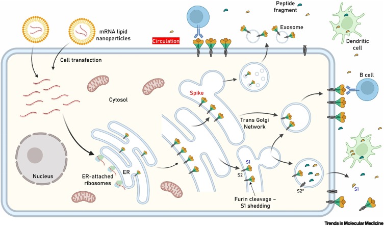
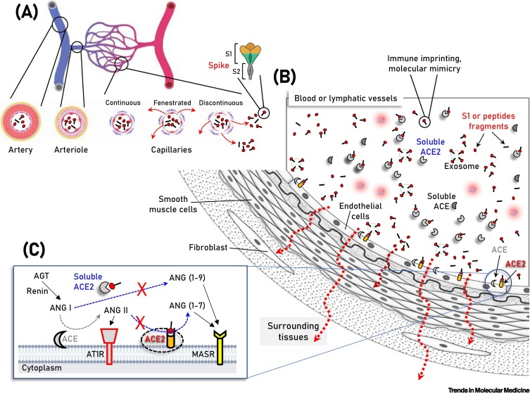
Vaccination is a major tool for mitigating the coronavirus disease 2019 (COVID-19) pandemic, and mRNA vaccines are central to the ongoing vaccination campaign that is undoubtedly saving thousands of lives. However, adverse effects (AEs) following vaccination have been noted which may relate to a proinflammatory action of the lipid nanoparticles used or the delivered mRNA (i.e., the vaccine formulation), as well as to the unique nature, expression pattern, binding profile, and proinflammatory effects of the produced antigens - spike (S) protein and/or its subunits/peptide fragments - in human tissues or organs. Current knowledge on this topic originates mostly from cell-based assays or from model organisms; further research on the cellular/molecular basis of the mRNA vaccine-induced AEs will therefore promise safety, maintain trust, and direct health policies.

Keywords: COVID-19; SARS-CoV-2; adverse effects; lipid nanoparticle; mRNA vaccine; spike protein.


Figures

Figure 1
Figure 1
Key figure. Antigen expression–localization following cell transfection with spike (S) protein mRNA-containing lipid nanoparticles (LNPs) used in anti-severe acute respiratory syndrome coronavirus 2 (SARS-CoV-2) mRNA vaccines. Following LNP internalization and mRNA release, the authentic viral signal peptide (as in the Pfizer–BioNTech and Moderna vaccines) drives antigen production in the lumen of the endoplasmic reticulum (ER) where it adopts its natural transmembrane localization via subunit 2 (S2) anchoring. After sorting in the trans Golgi network (TGN), S protein acquires its final position in the transfected human cell membrane, where S1 is exposed to the extracellular space (i.e., may face circulation). Although the extent of antigen expression per cell remains unknown, it is reasonable to assume that this process results in rather extended decoration of transfected cells with S protein. Furin-mediated proteolytic cleavage (as in SARS-CoV-2-infected cells) in the absence of a mutated S1/S2 furin cleavage site at the TGN may result in shedding of cleaved S1 and conversion of S2 into its postfusion structure (S2*). Antigen sorting and trafficking may also induce the release of S protein-containing exosomes. The events shown will occur in the apical and/or basolateral surfaces of polarized (e.g., epithelial) cells. The Pfizer–BioNTech and Moderna constructs do not contain a mutated S1/S2 furin cleavage site. Further research will clarify the impact of the S1/S2 subunits stabilizing D614G (or other) mutation or of a mutated furin cleavage site in antigen distribution, the immunogenicity of the vaccine, and induced adverse events (AEs). Also shown are dendritic cells (professional antigen-presenting cells, APCs) engulfing circulating antigens, and antibody-mediated binding of B cells to cell-anchored antigens.
Figure 2
Figure 2
Schematic of the vasculature components showing vaccination-produced S protein/subunits/peptide fragments in the circulation, as well as soluble or endothelial cell membrane-attached angiotensin-converting enzyme 2 (ACE2). (A,B) Parallel to immune system activation, circulating S protein/subunits/peptide fragments (B) binding to ACE2 may occur not only to ACE2-expressing endothelial cells, but also in multiple cell types of the vasculature and surrounding tissues due to antigen diffusion (e.g., in fenestrated or discontinuous capillary beds) (A, red arrows). These series of molecular events are unlikely for any severe acute respiratory syndrome coronavirus 2 (SARS-CoV-2)-related antigen in the absence of severe coronavirus disease 2019 (COVID-19), where SARS-CoV-2 is contained in the respiratory system. In (C) the two counteracting pathways of the renin–angiotensin system (RAS), namely the ‘conventional’ arm, that involves ACE which generates angiotensin II (ANG II) from angiotensin I (ANG I), and the ACE2 arm which hydrolyzes ANG II to generate angiotensin (1–7) [ANG (1–7)] or ANG I to generate angiotensin (1–9) [ANG (1–9)] are depicted. ANG II binding and activation of the ANG II type 1 receptor (AT1R) promotes inflammation, fibrotic remodeling, and vasoconstriction, whereas the ANG (1–7) and ANG (1–9) peptides binding to MAS receptor (MASR) activate antifibrotic, anti-inflammatory pathways and vasodilation. Additional modules of the RAS (i.e., renin and angiotensinogen, AGT) are also shown. Abbreviation: AT1R, angiotensin II type 1 receptor.

Comment in

Similar articles

Cited by

References

    1. Jin J., et al. Individual and community-level risk for COVID-19 mortality in the United States. Nat. Med. 2021;27:264–269. - PubMed
    1. O’Driscoll M., et al. Age-specific mortality and immunity patterns of SARS-CoV-2. Nature. 2021;590:140–145. - PubMed
    1. Gaebler C., et al. Evolution of antibody immunity to SARS-CoV-2. Nature. 2021;591:639–644. - PMC - PubMed
    1. Andersson M.I., et al. SARS-CoV-2 RNA detected in blood products from patients with COVID-19 is not associated with infectious virus. Wellcome Open Res. 2020;5:181. - PMC - PubMed
    1. Wang W., et al. Detection of SARS-CoV-2 in different types of clinical specimens. JAMA. 2020;323:1843–1844. - PMC - PubMed