. 2022 May 11;8(1):258.
doi: 10.1038/s41420-022-01046-w.

Mitochondrial damage and activation of the cytosolic DNA sensor cGAS-STING pathway lead to cardiac pyroptosis and hypertrophy in diabetic cardiomyopathy mice

Affiliations

Mitochondrial damage and activation of the cytosolic DNA sensor cGAS-STING pathway lead to cardiac pyroptosis and hypertrophy in diabetic cardiomyopathy mice

Meiling Yan et al. Cell Death Discov. .

Abstract

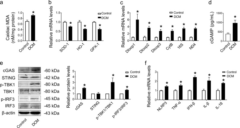
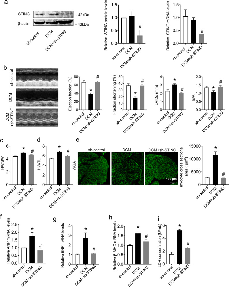
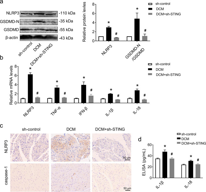
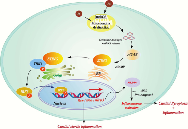
Diabetic cardiomyopathy (DCM) is a serious cardiac complication of diabetes that currently lacks specific treatment. The cyclic GMP-AMP synthase (cGAS)-stimulator of interferon genes (STING) signaling pathway has been suggested to contribute to the pathogenesis of cardiovascular diseases. However, whether cGAS-STING is involved in the development of DCM has not been established. Our study aimed to determine the role of cGAS-STING in the initiation of nucleotide-binding oligomerization domain-like receptor pyrin domain containing 3 (NLRP3) inflammasome-induced cardiac pyroptosis and chronic inflammation during the pathogenesis of DCM. C57BL/6J mice were preinjected with adeno-associated virus 9 (AAV9) intravenously via the tail vein to specifically knock down myocardial STING. After four weeks, mice with myocardium-specific knockdown of STING received injections of streptozotocin (STZ; 50 mg/kg) and a high-fat diet to induce diabetes. Measurements included echocardiography, immunohistochemical analyses, wheat germ agglutinin (WGA) staining, and western blotting. Here, we showed that the cGAS-STING signaling pathway was activated in diabetic hearts, which was indicated by the increased phosphorylation of TANK-binding kinase 1 (TBK1) and interferon (IFN) regulatory factor 3 (IRF3), leading to the activation of the NLRP3 inflammasome in the hearts of diabetic mice and proinflammatory cytokine release into serum. Moreover, STING knockdown via adeno-associated virus-9 (AAV9) in diabetic mouse heart alleviated cardiac pyroptosis and the inflammatory response, prevented diabetes-induced hypertrophy, and restored cardiac function. Mechanistically, we showed that palmitic acid (PA)-induced lipotoxicity impairs mitochondrial homeostasis, producing excessive mitochondrial reactive oxygen species (mtROS), which results in oxidative damage to mitochondrial DNA (mtDNA) and its release into the cytoplasm while switching on cGAS-STING-mediated pyroptosis in cardiomyocytes, thereby worsening the progression of diabetic cardiomyopathy. Our study demonstrated that activation of the cGAS-STING pathway caused by mitochondrial oxidative damage and mtDNA escape induced by free fatty acids promoted pyroptosis and proinflammatory responses in cardiomyocytes in a NLRP3 inflammasome-dependent manner, thus promoting myocardial hypertrophy during the progression of DCM.


Conflict of interest statement

The authors declare that the research was conducted in the absence of any commercial or financial relationships that could be construed as a potential conflict of interest.

Figures

Fig. 1
Fig. 1. Oxidative stress-induced leakage of mtDNA and activation of the cGAS-STING pathway accompanied with inflammation in DCM mice hearts.
a Assessment of the level of MDA in fresh heart tissues. n = 3 in each group. b mRNA levels of SOD-1, HO-1, and GPX-1 determined in tissues by qRT–PCR. n = 3 in each group. c mRNA levels of Dloop1, Dloop2, Dloop3, CytB, 16S, and ND4 determined in tissues by qRT–PCR. n = 3 in each group. d The contents of cGAMP in the hearts of DCM mice. n = 7 in each group. e Representative protein levels of cGAS, STING, p-TBK1/TBK1, and p-IRF3/IRF3 normalized to β-actin in heart tissues by western blot. n = 3 in each group. f mRNA levels of NLRP3, TNF-α, IFN-β, IL-1β, and IL-18 in tissues. n = 3 in each group. The results were normalized to β-actin. Values are the mean ± SEM. *P < 0.05 vs. the control group. DCM diabetic cardiomyopathy, MDA malondialdehyde.
Fig. 2
Fig. 2. Knockdown of STING significantly improved cardiac function and inhibited cardiac hypertrophy in DCM mice.
a Protein and mRNA levels of STING normalized to β-actin in heart tissues. n = 3 in each group. b Transthoracic echocardiography was performed to observe changes in cardiac function and morphology in both sh-control and DCM animals with or without shRNA of STING. Statistics of ejection fraction, fractional shortening, LVIDs and peak E to peak A (E/A) ratio. n = 7 in each group. c, d Statistical results of heart weight/body weight (HW/BW) and heart weight/tibia length (HW/TL). n = 6 in each group. e Quantitative analysis of myocyte cross-sectional area by WGA staining. n = 3 in each group. fh mRNA levels of ANP, BNP, and β-MHC in heart tissues. n = 3 in each group. i LDH release in each group. n = 6 in each group. Values are the mean ± SEM. *P < 0.05 vs. the sh-control group, #P < 0.05 vs. the DCM group. DCM diabetic cardiomyopathy, sh-STING shRNA of STING.
Fig. 3
Fig. 3. Knockdown of STING in DCM mice attenuated cardiomyocyte pyroptosis and inflammation.
a Representative western blot analysis of NLRP3 and GSDMD-N/GSDMD in sh-control and DCM hearts after treatment with or without shRNA of STING and normalized to β-actin. n = 3 in each group. b mRNA levels of NLRP3, TNF-α, IFN-β, IL-1β, and IL-18 in heart tissues. n = 3 in each group. c Immunohistochemical staining analysis of NLRP3 and caspase-1 in heart tissues. n = 3 in each group. d Levels of IL-1β and IL-18 in serum analyzed by ELISA. n = 5 in each group. Values are the mean ± SEM. *P < 0.05 vs. the sh-control group, #P < 0.05 vs. the DCM group. DCM diabetic cardiomyopathy, shRNA of STING, adeno-associated virus 9-sh-STING.
Fig. 4
Fig. 4. PA activated cGAS-STING pathway in cardiomyocyte by induced mtDNA release and oxidative damage, leading to cell death.
a Effect of PA on cardiomyocyte viability. H9C2 cells were incubated with different concentrations (50, 100, 200, 300, 400, or 500 μM) of PA for 24 h and then assayed by cell counting kit-8. n = 5 in each group. b Representative flow cytometry image and quantitative analysis of the intracellular mtROS levels. n = 5 in each group. c Representative images of tetrachloro-tetraethyl benzimidazole carbocyanine iodide (JC-1) staining in NRCMs. n = 5 in each group. d mtDNA levels of Dloop1, Dloop2, Dloop3, CytB, 16S, and ND4 after treatment with or without PA in NRCMs. n = 5 in each group. e Oxidative guanine base damage of DNA was detected by fluorescence-based immunocytochemistry using specific antibodies for 8-OHdG in NRCMs. n = 6 in each group. f Western blot bands and analysis of cGAS, STING, p-TBK1/TBK1, and p-IRF3/IRF3 after treatment with PA with or without EtBr in H9C2 cells normalized to β-actin. n = 3 in each group. g The mRNA levels of NLRP3, TNF-α, IFN-β, IL-1β, and IL-18 in H9C2 cells. n = 3 in each group. h Representative flow cytometry image and the corresponding quantification showing the number of PI+ cells in the pyroptosis population. Hoechst 33342/PI double staining was performed by flow cytometry to detect pyroptosis in H9C2 cells. n = 6 in each group. i LDH release from each group of H9C2 cells. n = 9 in each group. Values are the mean ± SEM. *P < 0.05 vs. the control group, #P < 0.05 vs. PA-treated H9C2 cells. PA palmitic acid.
Fig. 5
Fig. 5. Inhibited cGAS-STING pathway effectively reduced cardiomyocyte pyroptosis and hypertrophy induced by PA treatment.
a Western blot analysis of NLRP3 and GSDMD-N/GSDMD after treatment with PA with or without siRNA of cGAS or siRNA of STING in H9C2 cells normalized to β-actin. n = 3 in each group. b Relative mRNA levels of NLRP3, TNF-α, IFN-β, IL-1β, and IL-18 in H9C2 cells. n = 3 in each group. c Representative flow cytometry image and the corresponding quantification showing the number of PI+ cells as the pyroptosis population. n = 4 in each group. d LDH release from each group of H9C2 cells. n = 8 in each group. e H9C2 cells were incubated with PA with or without siRNA of cGAS or siRNA of STING for 24 h and then evaluated by cell counting kit-8. n = 8 in each group. f, g Representative images and quantitative analysis of caspase-1 (red) and TUNEL (green) double-positive cells in PA-induced NMCMs. n = 5 in each group. h, i Quantitative analysis of the myocyte cross-sectional area by α-actinin staining in NMCMs. n = 6 in each group. j mRNA levels of ANP, BNP, and β-MHC in H9C2 cells. n = 3 in each group. Values are the mean ± SEM. *P < 0.05 vs. NC, #P < 0.05 vs. PA-treated cells. NC negative control, PA palmitic acid, si-cGAS small interfering RNA of cGAS, si-STING small interfering RNA of STING.
Fig. 6
Fig. 6. Proposed mechanism of PA-induced myocardial pyroptosis mediated by cGAS-STING pathway activation in DCM.
PA causes mitochondrial damage and the release of mtDNA into the cytosol, which is converted to cGAMP by cGAS. cGAMP activates STING, which facilitates TBK-1-mediated IRF3 phosphorylation. Phosphorylated IRF3 enters the nucleus and induces type I interferon expression and NLRP3 inflammasome activation, which leads to the exacerbation of myocardial inflammation and pyroptosis.

References

    1. Dillmann WH. Diabetic cardiomyopathy. Circ Res. 2019;124:1160–2. - PMC - PubMed
    1. Gilbert RE, Krum H. Heart failure in diabetes: effects of anti-hyperglycaemic drug therapy. Lancet. 2015;385:2107–17. - PubMed
    1. Borghetti G, Lewinski DV, Eaton DM, Sourij H, Wallner M. Diabetic cardiomyopathy: current and future therapies. Beyond glycemic control. Front Physiol. 2018;9:1514. - PMC - PubMed
    1. Pedersen PL. Mitochondrial matters of the heart: a plethora of regulatory modes to maintain function for a long lifetime. J Bioenerg Biomembranes. 2009;41:95–98. - PubMed
    1. Nakamura M, Sadoshima J. Cardiomyopathy in obesity, insulin resistance or diabetes. J Physiol. 2019;598:2977–93. - PubMed