. 2022 May 10;14(1):25.
doi: 10.1038/s41368-022-00174-4.

FAM64A promotes HNSCC tumorigenesis by mediating transcriptional autoregulation of FOXM1

Affiliations

FAM64A promotes HNSCC tumorigenesis by mediating transcriptional autoregulation of FOXM1

Xinyuan Zhao et al. Int J Oral Sci. .

Abstract

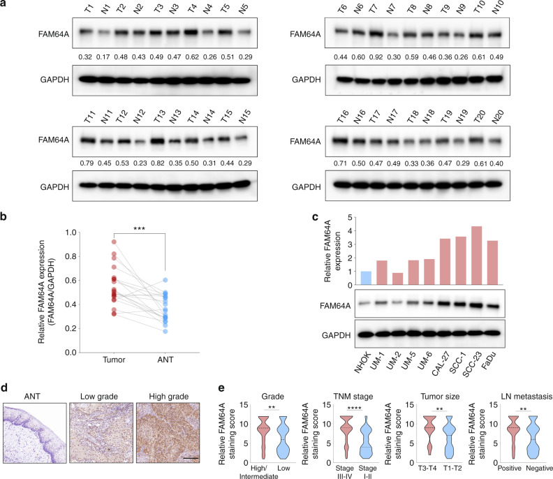
Head and neck squamous cell carcinoma (HNSCC) still lacks effective targeted treatment. Therefore, exploring novel and robust molecular targets is critical for improving the clinical outcome of HNSCC. Here, we reported that the expression levels of family with sequence similarity 64, member A (FAM64A) were significantly higher in HNSCC tissues and cell lines. In addition, FAM64A overexpression was found to be strongly associated with an unfavorable prognosis of HNSCC. Both in vitro and in vivo evidence showed that FAM64A depletion suppressed the malignant activities of HNSCC cells, and vice versa. Moreover, we found that the FAM64A level was progressively increased from normal to dysplastic to cancerous tissues in a carcinogenic 4-nitroquinoline-1-oxide mouse model. Mechanistically, a physical interaction was found between FAM64A and forkhead box protein M1 (FOXM1) in HNSCC cells. FAM64A promoted HNSCC tumorigenesis not only by enhancing the transcriptional activity of FOXM1, but also, more importantly, by modulating FOXM1 expression via the autoregulation loop. Furthermore, a positive correlation between FAM64A and FOXM1 was found in multiple independent cohorts. Taken together, our findings reveal a previously unknown mechanism behind the activation of FOXM1 in HNSCC, and FAM64A might be a promising molecular therapeutic target for treating HNSCC.


Conflict of interest statement

The authors declare no competing interests.

Figures

Fig. 1
Fig. 1
Clinical significance of FAM64A in HNSCC. a, b Western blot analysis of FAM64A protein expression in 20 HNSCC tumor tissues and paired ANTs. c Western blot analysis of FAM64A protein expression in HNSCC cell lines and NHOK. d, e IHC analysis of FAM64A staining intensity in HNSCC tissues and nontumor tissues. Data are presented as the mean ± SD. **P < 0.01, ***P < 0.001, ****P < 0.0001. Scale bar: 200 μm for (d)
Fig. 2
Fig. 2
FAM64A promotes the malignant behaviors of HNSCC cells in vitro and in vivo. a Validation via western blotting of the depletion effect of shFAM64A in SCC-1 cells. b FAM64A depletion significantly reduced the percentage of EdU-positive cells and suppressed the tumor sphere formation capability and colony formation capacity of SCC-1 cells. c, d The proliferation, tumor sphere formation, and colony formation capacities were significantly enhanced in FAM64A overexpressing cells compared to control cells. e, f In vivo xenograft studies demonstrated that FAM64A depletion significantly attenuated the size, weight, and volume of tumors derived from SCC-1 cells. g, h Ectopic expression of FAM64A dramatically increased the size, weight, and volume of tumors derived from SCC-1 cells. Data are presented as the mean ± SD. ***P < 0.001, ****P < 0.0001. Scale bar: 50 μm for upper (b) and (d) and 200 μm for lower (b) and (d)
Fig. 3
Fig. 3
FAM64A interacts with FOXM1, and regulates FOXM1 expression via the FOXM1 autoregulation loop. a FAM64A-binding proteins were revealed by immunoblotting and silver staining. b, c Co-IP assays were performed to further validate the interaction between FAM64A and FOXM1 in HNSCC cells. d, e FAM64A depletion reduced FOXM1 protein levels in both HNSCC cell lines, and ectopic expression of FAM64A increased FOXM1 levels in SCC-1 and SCC-23 cells. f, g SCC-1 cells/SCC-23 cells with or without FAM64A depletion were treated with CHX for different periods of time, and the expression level of FOXM1 was detected by immunoblotting. h FAM64A upregulation enhanced the expression level of FOXM1 mRNA in SCC-1 cells, and FOXM1 downregulation abrogated these effects. i FAM64A depletion reduced the FOXM1 mRNA level in HNSCC cells. j CHIP-qPCR results demonstrated that FAM64A depletion significantly reduced the occupancy of FOXM1 on the promoter of FOXM1 in HNSCC cells. k FAM64A enhanced FOXM1 transcriptional activity on the FOXM1 promoter in a dose-dependent fashion. l FAM64A depletion decreased FOXM1 transcriptional activity on the FOXM1 promoter in HNSCC cells. Data are presented as the mean ± SD. **P < 0.01, ***P < 0.001, ns=not significant
Fig. 4
Fig. 4
FAM64A regulates FOXM1 transcriptional activity in HNSCC cells. a FAM64A was found to be positively correlated with FOXM1 target genes including CCNB1, CCNB2, BIRC5, AURKA, AURKB, XRCC1, SOX2, and PLK1 in TCGA HNSCC cohort. b qRT-PCR results revealed that FAM64A overexpression enhanced the expression levels of FOXM1 target genes in SCC-1 cells, and FOXM1 depletion abrogated these promotive effects. c FAM64A depletion suppressed the expression levels of FOXM1 target genes in SCC-1 cells. d CHIP-qPCR demonstrated that FAM64A downregulation markedly reduced the occupancy of FOXM1 on the promoters of CCNB1, CCNB2, BIRC5, AURKA, AURKB, XRCC1, SOX2, and PLK1 in SCC-1 cells. Data are presented as the mean ± SD. *P < 0.05, **P < 0.01, ***P < 0.001
Fig. 5
Fig. 5
FOXM1 overexpression rescued the tumor suppressive effects of FAM64A depletion in vitro and in vivo. a Western blot analysis of FOXM1 and FAM64A protein expression in SCC-1 cells with the indicated modifications. b, c The proliferation, tumor sphere formation and colony formation capacities of SCC-1 cells were suppressed by FAM64A depletion. These suppressive effects were reversed by ectopic expression of FOXM1. d, e FAM64A depletion repressed the growth of tumors derived from SCC-1 cells, and the inhibitory effects were rescued by FOXM1 overexpression. f The Ki-67, CD44, SOX2, and FOXM1 staining intensity were decreased in xenograft tumor tissues formed by shFAM64A-treated SCC-1 cells, and FOXM1 overexpression abrogated these effects. Data are presented as the mean ± SD. **P < 0.01, ***P < 0.001. Scale bar: 50 μm for upper (b) and (f), 200 μm for lower (b)
Fig. 6
Fig. 6
FAM64A expression is positively correlated with FOXM1 expression in HNSCC tumor tissues and the prognostic value of the FAM64A-FOXM1 axis in HNSCC. a, b The expression level of FAM64A in HNSCC tumor tissues was positively correlated with FOXM1 expression. c HNSCC patients in the high FAM64A IHC staining score group suffered worse OS than those in the low FAM64A IHC staining score group. d Patients in the high FOXM1 IHC staining score group had more unfavorable OS than those in the low FOXM1 IHC staining score group. e, f Univariate analysis revealed that FAM64A staining score was strongly associated with HNSCC patient OS, and the multivariate analysis demonstrated that it was an independent prognostic factor for HNSCC. Scale bar: 200 μm for (a)
Fig. 7
Fig. 7
A graphical model for FAM64A-mediated tumorigenesis in HNSCC. FAM64A promotes HNSCC tumorigenesis by increasing FOXM1 transcriptional activity and activating FOXM1 expression via the FOXM1 autoregulation loop.

References

    1. Leemans CR, Snijders PJF, Brakenhoff RH. The molecular landscape of head and neck cancer. Nat. Rev. Cancer. 2018;18:269–282. doi: 10.1038/nrc.2018.11. - DOI - PubMed
    1. Jiang Z, et al. Research on neck dissection for oral squamous-cell carcinoma: a bibliometric analysis. Int. J. Oral. Sci. 2021;13:13. doi: 10.1038/s41368-021-00117-5. - DOI - PMC - PubMed
    1. Banerjee R, et al. Phosphorylation of TRIP13 at Y56 induces radiation resistance but sensitizes head and neck cancer to cetuximab. Mol. Ther. 2022;30:468–484. doi: 10.1016/j.ymthe.2021.06.009. - DOI - PMC - PubMed
    1. Li Q, et al. Prognostic value of tertiary lymphoid structure and tumour infiltrating lymphocytes in oral squamous cell carcinoma. Int. J. Oral. Sci. 2020;12:24. doi: 10.1038/s41368-020-00092-3. - DOI - PMC - PubMed
    1. Johnson DE, et al. Head and neck squamous cell carcinoma. Nat. Rev. Dis. Prim. 2020;6:92. doi: 10.1038/s41572-020-00224-3. - DOI - PMC - PubMed

Publication types