

Development of a standardized in vitro model to reproduce hydrophilic acrylic intraocular lens calcification
- PMID: 35538104
- PMCID: PMC9090772
- DOI: 10.1038/s41598-022-11486-0
Development of a standardized in vitro model to reproduce hydrophilic acrylic intraocular lens calcification
Abstract
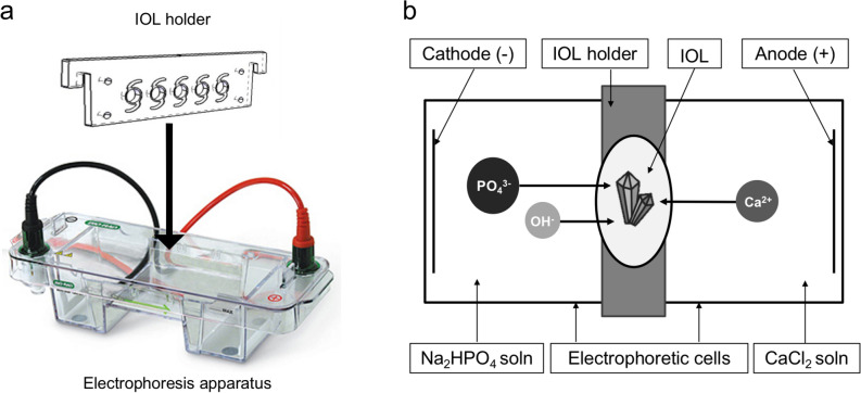
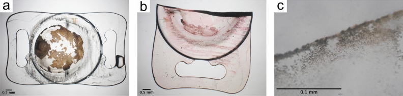
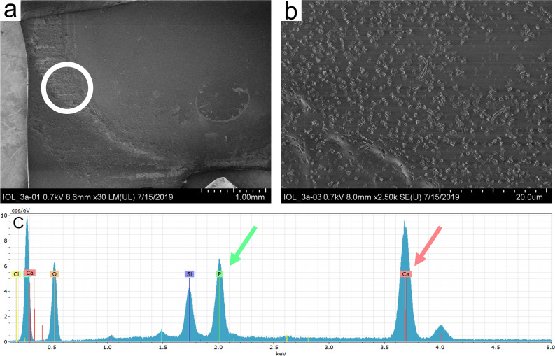
Opacification through calcification of hydrophilic acrylic intraocular lenses (IOL) is a severe complication after cataract surgery. Causing symptoms that range from glare through to severe vision loss, the only effective therapy is explantation of the opacified IOL so far. Although IOL calcification is a well-described phenomenon, its pathogenesis is not fully understood yet. The purpose of the current study was to develop a laboratory model to replicate IOL calcification. Calcification could be reproduced using a horizontal electrophoresis and aqueous solutions of calcium chloride and disodium hydrogen phosphate. The analysis of the in vitro calcified IOLs was performed using light microscopy, Alizarin Red and Von Kossa staining, scanning electron microscopy, energy dispersive x-ray spectroscopy and electron crystallography using transmission electron microscopy and electron diffraction. The presented laboratory model could be used to identify hydrophilic IOLs that are at risk to develop calcification and to assess the influence of associated risk factors. In addition, it can serve as a research tool to further understand this pathology.
© 2022. The Author(s).
Conflict of interest statement
The authors declare no competing interests.
Figures


Similar articles
-
Localized calcification of hydrophilic acrylic intraocular lenses after posterior segment procedures.J Cataract Refract Surg. 2019 Dec;45(12):1801-1807. doi: 10.1016/j.jcrs.2019.07.015. J Cataract Refract Surg. 2019. PMID: 31856993
-
Dense opacification of the optical component of a hydrophilic acrylic intraocular lens: a clinicopathological analysis of 9 explanted lenses.J Cataract Refract Surg. 2001 Sep;27(9):1485-92. doi: 10.1016/s0886-3350(01)00841-0. J Cataract Refract Surg. 2001. PMID: 11566535
-
Hydrophilic acrylic intraocular lens optic and haptics opacification in a diabetic patient: bilateral case report and clinicopathologic correlation.Ophthalmology. 2002 Nov;109(11):2042-51. doi: 10.1016/s0161-6420(02)01265-4. Ophthalmology. 2002. PMID: 12414413
-
Microscopic Characteristics of Late Intraocular Lens Opacifications.Arch Pathol Lab Med. 2021 Jun 1;145(6):759-767. doi: 10.5858/arpa.2019-0626-RA. Arch Pathol Lab Med. 2021. PMID: 33091924 Review.
-
Complications of cataract and refractive surgery: a clinicopathological documentation.Trans Am Ophthalmol Soc. 2001;99:95-107; discussion 107-9. Trans Am Ophthalmol Soc. 2001. PMID: 11797325 Free PMC article. Review.
Cited by
-
How do intraocular lens materials influence the outcome of cataract surgery?Curr Opin Ophthalmol. 2025 Jan 1;36(1):18-24. doi: 10.1097/ICU.0000000000001095. Epub 2024 Oct 23. Curr Opin Ophthalmol. 2025. PMID: 39446645 Free PMC article. Review.
-
[Hydrophobic surface properties of hydrophilic acrylic lenses do not protect against calcification].Ophthalmologie. 2023 Oct;120(10):1022-1028. doi: 10.1007/s00347-023-01862-0. Epub 2023 May 12. Ophthalmologie. 2023. PMID: 37171476 German.
-
Imaging Function and Relative Light Transmission of Explanted Opacified Hydrophilic Acrylic Intraocular Lenses.Diagnostics (Basel). 2023 May 19;13(10):1804. doi: 10.3390/diagnostics13101804. Diagnostics (Basel). 2023. PMID: 37238287 Free PMC article.
-
Impact of Calcium and Phosphorus Levels on Optical Deterioration in Primary and Secondary Intraocular Lens Calcification.Transl Vis Sci Technol. 2024 Oct 1;13(10):18. doi: 10.1167/tvst.13.10.18. Transl Vis Sci Technol. 2024. PMID: 39388178 Free PMC article.
-
Higher phosphate concentrations as in aqueous humor of diabetic patients increase intraocular lens calcification.BMC Ophthalmol. 2024 Aug 23;24(1):363. doi: 10.1186/s12886-024-03553-z. BMC Ophthalmol. 2024. PMID: 39179956 Free PMC article.
References
Publication types
MeSH terms
LinkOut - more resources
Full Text Sources
