. 2022 May 10;12(1):7684.
doi: 10.1038/s41598-022-11699-3.

A non-canonical Raf function is required for dorsal-ventral patterning during Drosophila embryogenesis

Affiliations

A non-canonical Raf function is required for dorsal-ventral patterning during Drosophila embryogenesis

Jay B Lusk et al. Sci Rep. .

Abstract

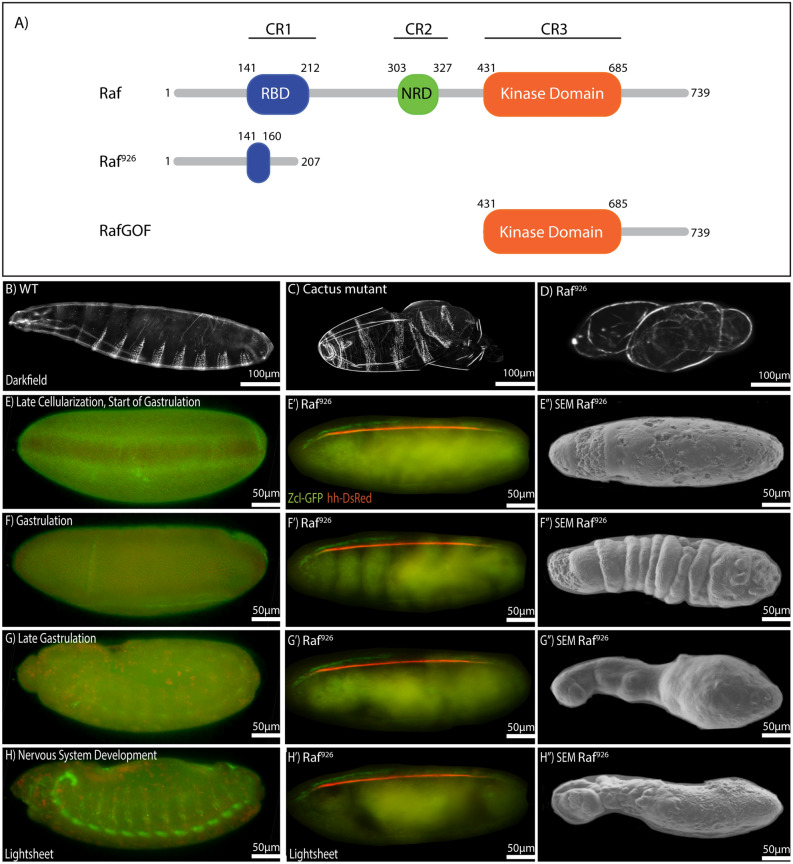
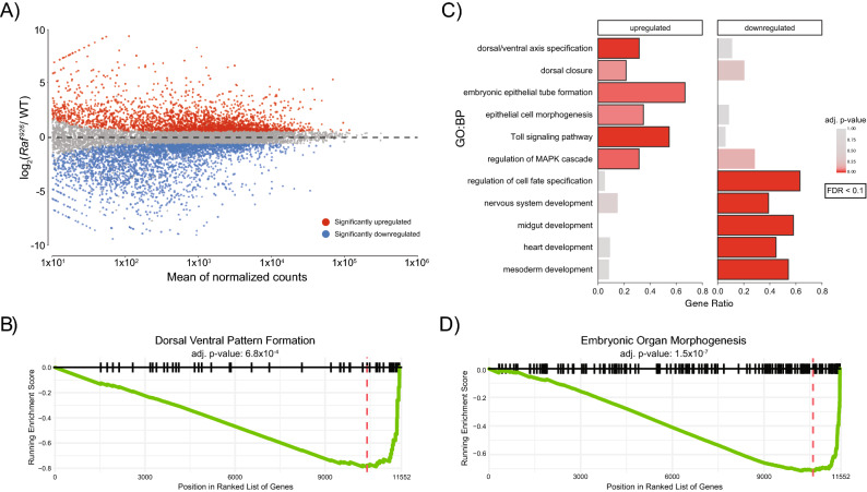
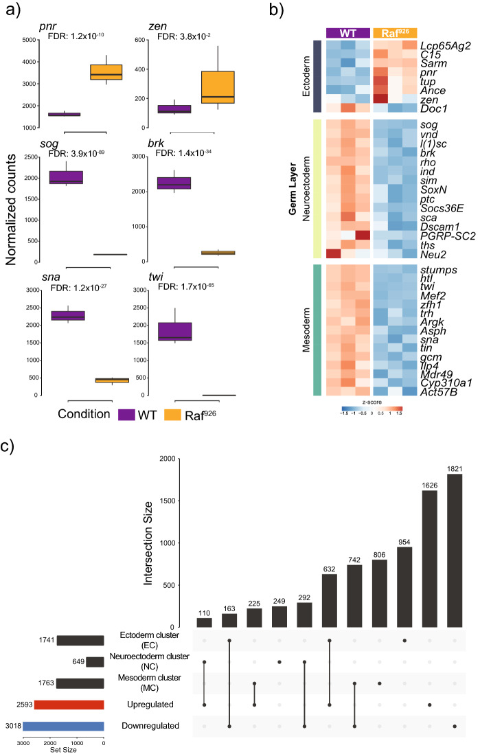
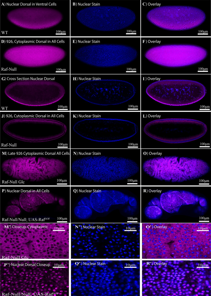
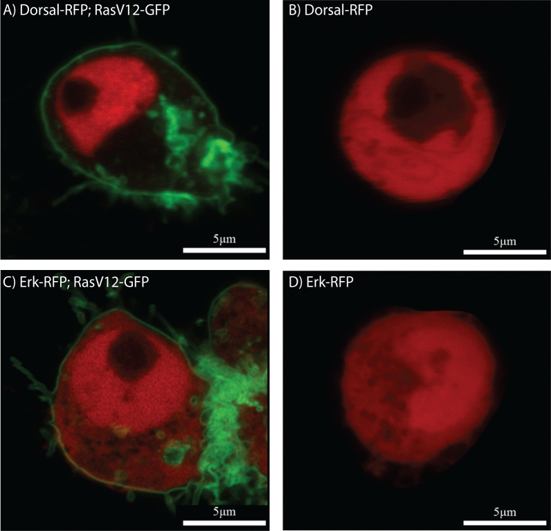
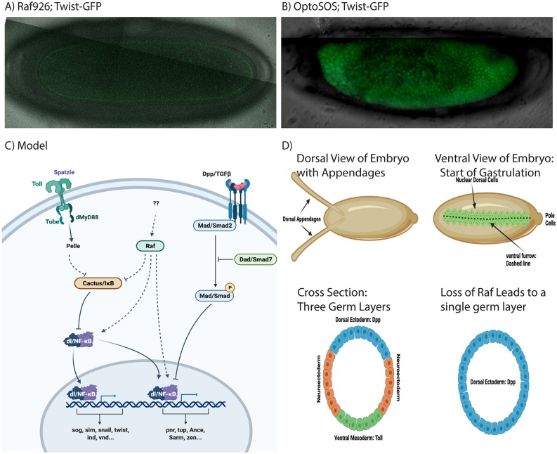
Proper embryonic development requires directional axes to pattern cells into embryonic structures. In Drosophila, spatially discrete expression of transcription factors determines the anterior to posterior organization of the early embryo, while the Toll and TGFβ signalling pathways determine the early dorsal to ventral pattern. Embryonic MAPK/ERK signaling contributes to both anterior to posterior patterning in the terminal regions and to dorsal to ventral patterning during oogenesis and embryonic stages. Here we describe a novel loss of function mutation in the Raf kinase gene, which leads to loss of ventral cell fates as seen through the loss of the ventral furrow, the absence of Dorsal/NFκB nuclear localization, the absence of mesoderm determinants Twist and Snail, and the expansion of TGFβ. Gene expression analysis showed cells adopting ectodermal fates much like loss of Toll signaling. Our results combine novel mutants, live imaging, optogenetics and transcriptomics to establish a novel role for Raf, that appears to be independent of the MAPK cascade, in embryonic patterning.


Conflict of interest statement

The authors declare no competing interests.

Figures

Figure 1
Figure 1
Schematic illustration, cuticular phenotype and developmental progression of Raf926 mutant embryo. (A) Domain organization of Drosophila Raf, Raf926 and RafGOF. Raf protein contains the Ras-binding domain (RBD) in conserved region 1 (CR1), negative regulatory domain (NRD) in CR2 and protein kinase domain in CR3, with a total length of 739 amino acids. Raf926 comprised of an altered RBD with 207 amino acids in total due to a deletion of 17 nucleotides which resulted in a frameshift and premature stop codon. RafGOF contains the kinase domain only, with total length of 309 amino acids. Dark field micrographs of the cuticles of wild type embryo showing normal distribution of ventral denticles and dorsal hair (B), a cactus1 mutant embryo showing strongly ventralized phenotype with ventral denticles expression among the dorsal hair (C) and Raf926 mutant embryo showing a strongly dorsalized phenotype with elongated, tube-like, twisted body entirely covered by dorsal hair (D). (EH) Still images of developmental stages of a wild-type embryo to be compared to still images of developmental stages of Raf926 embryo from lightsheet (E′H′) and scanning electron microscope (SEM) (E″H″). Raf926 embryos develop normally up to cellularization stage. Defective gastrulation is characterized by frequent twisting, elongation of twisted segments, fusions of segments into three main sacs, followed by tissue death. Embryo was visualised with Utrophin-GFP and Histone-RFP, with anterior to the left. Stills were extracted from Video 3.
Figure 2
Figure 2
Transcriptional profiling of WT, Raf926 embryos identifies significant differences in the expression of genes involved in Dorsal/Ventral patterning, organogenesis and the Toll pathway. (A) MA-plot shows a large number of genes are significantly differentially expressed between Raf926 mutant and WT embryos (three biological replicates per condition). (B) GSEA identifies that genes annotated as involved in dorsal–ventral pattern formation are altered in Raf926 mutants. (C) GO Biological Process (GO:BP) enrichment analysis identifies that the significant up- and downregulated genes are enriched for distinct processes. (D) GSEA identifies that genes are involved in embryonic organ morphogenesis and perturbed in Raf926 mutants.
Figure 3
Figure 3
Comparison of expression changes in observed Raf926 mutants with key germ layer marker genes. (A) Expression profiles of key germ layer marker genes in WT and Raf926 mutant embryos. (B) Raf926 mutants show upregulation of ectoderm marker genes, along with downregulation of neuroectoderm and mesoderm marker genes. (C) Upset plot showing the overlap between differentially expressed genes in Raf926 mutant with clusters of gene expression changes observed after generating embryos with only one germ layer after perturbing the Toll pathway (see S. Fig. 3D).
Figure 4
Figure 4
Dorsal nuclear localization in WT, Raf926 embryo and Raf926 embryo expressing RafGOF. Surface view of the lateral orientation of the wildtype embryo (A) undergoing cellularization with a nuclear Dorsal gradient along the dorsal–ventral axis, (B) shows the corresponding nuclear stain and (C) a merged image. Surface view of the lateral orientation of the Raf926 embryo (D) undergoing cellularization with complete exclusion of Dorsal from the nuclei. (E) The corresponding nuclear stain and (F) a merged image of both. Cross-sectional view of the lateral orientation of the (G) wildtype embryo showing the gradient nuclear localization of Dorsal along the dorsal–ventral axis, (H) shows the corresponding nuclear stain and (I) a merged image of both. Cross-sectional view of Raf926 embryo (J) showing the complete exclusion of Dorsal from the nucleus, (K) showing the corresponding nuclear stain and (L) a merged image of both. Surface view of (M, M′) Raf926 later stage embryo showing abnormality in development after cellularization and exclusion of Dorsal from the nucleus. The corresponding nuclear stain (N, N′) and merged images (O, O′). (P, P′) Raf926 embryo expressing RafGOF showing dorsal nuclear localization. (Q, Q′) shows the corresponding nuclear stain and (R, R′) the merged image.
Figure 5
Figure 5
Dorsal and Erk localization in vitro. (A) Nuclear localization of Dorsal (red) in S2R+ cell expressing the gain of function RasV12 (green). (B) Nuclear exclusion of Dorsal in S2R+ cell in the absence of RasV12 expression. Ubiquitous expression of Erk (red) in both cytoplasm and nucleus of S2R+ cell both (C) in the presence and (D) absence of RasV12 (green). The black spot in the nucleus in (AC) is the nucleolus.
Figure 6
Figure 6
Opto-SOS drives Twist nuclear localization. (A) Live, confocal imaging of Twist-GFP expression in a Raf926 embryo shows no expression. (B) Activation of Raf using Opto-SOS showed uniform Twist-GFP (C). A model for Raf in Dorsal/Ventral signaling. (D) Schematic representation of the Dorsal and Ventral sides of Drosophila embryos with dorsal appendages and ventral furrow cells highlighted followed by cross section views of the early germ layers as defined in wildtype and Raf mutants. Figures were drawn on Biorender.com.

Similar articles

Cited by

References

    1. Francois V, Solloway M, O'Neill JW, Emery J, Bier E. Dorsal-ventral patterning of the Drosophila embryo depends on a putative negative growth factor encoded by the short gastrulation gene. Genes Dev. 1994;8:2602–2616. doi: 10.1101/gad.8.21.2602. - DOI - PubMed
    1. Anderson KV, Bokla L, Nusslein-Volhard C. Establishment of dorsal-ventral polarity in the Drosophila embryo: The induction of polarity by the Toll gene product. Cell. 1985;42:791–798. doi: 10.1016/0092-8674(85)90275-2. - DOI - PubMed
    1. Anderson KV, Jurgens G, Nusslein-Volhard C. Establishment of dorsal-ventral polarity in the Drosophila embryo: Genetic studies on the role of the Toll gene product. Cell. 1985;42:779–789. doi: 10.1016/0092-8674(85)90274-0. - DOI - PubMed
    1. Von Ohlen T, Doe CQ. Convergence of dorsal, dpp, and egfr signaling pathways subdivides the drosophila neuroectoderm into three dorsal-ventral columns. Dev. Biol. 2000;224:362–372. doi: 10.1006/dbio.2000.9789. - DOI - PubMed
    1. Padgett RW, St Johnston RD, Gelbart WM. A transcript from a Drosophila pattern gene predicts a protein homologous to the transforming growth factor-beta family. Nature. 1987;325:81–84. doi: 10.1038/325081a0. - DOI - PubMed

Publication types

MeSH terms

Substances

LinkOut - more resources