. 2022 May 10;5(1):433.
doi: 10.1038/s42003-022-03400-1.

Nuclear soluble cGAS senses double-stranded DNA virus infection

Affiliations

Nuclear soluble cGAS senses double-stranded DNA virus infection

Yakun Wu et al. Commun Biol. .

Abstract

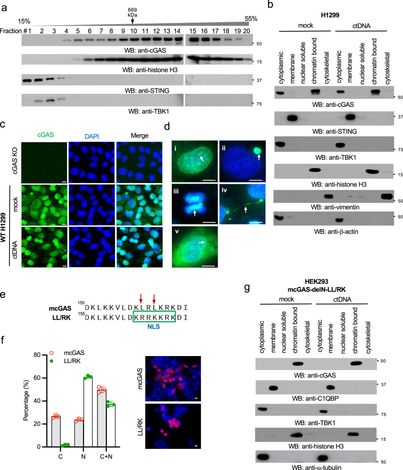
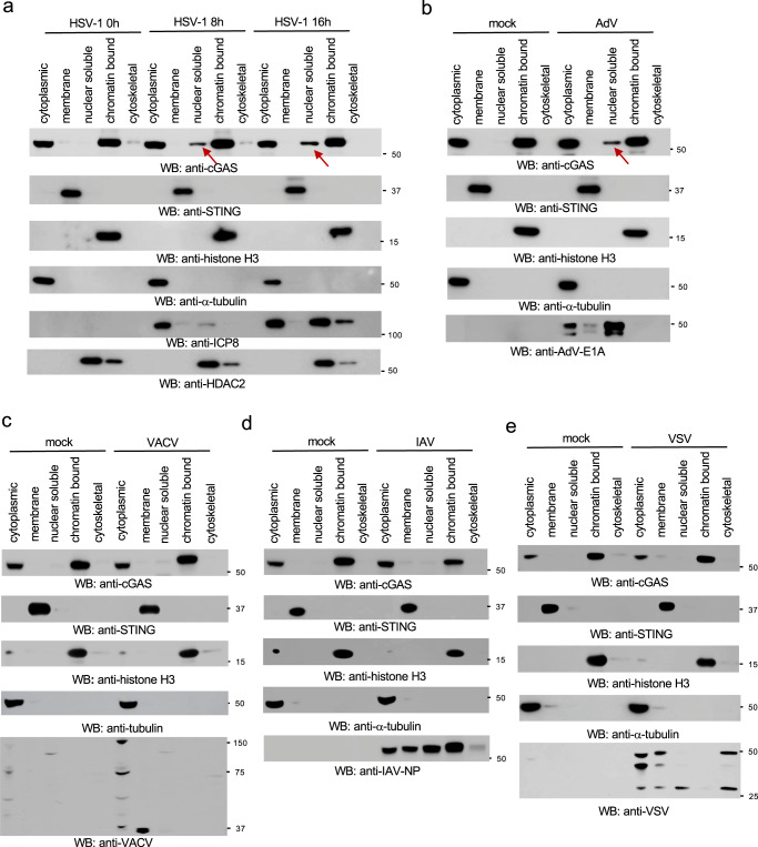
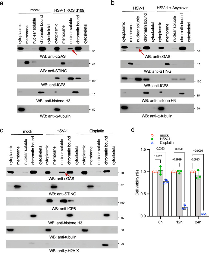
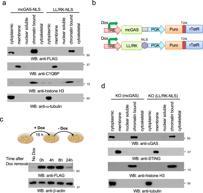
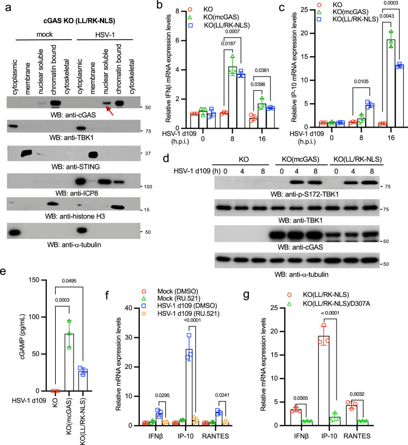
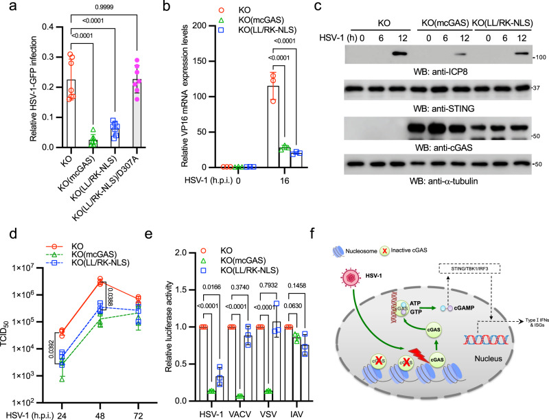
The DNA sensor cGAS detects cytosolic DNA and instigates type I interferon (IFN) expression. Recent studies find that cGAS also localizes in the nucleus and binds the chromatin. Despite the mechanism controlling nuclear cGAS activation is well elucidated, whether nuclear cGAS participates in DNA sensing is unclear. Here, we report that herpes simplex virus 1 (HSV-1) infection caused the release of cGAS from the chromatin into the nuclear soluble fraction. Like its cytosolic counterpart, the leaked nuclear soluble cGAS also sensed viral DNA, produced cGAMP, and induced mRNA expression of type I IFN and interferon-stimulated genes. Consistently, the nuclear soluble cGAS limited HSV-1 infection. Furthermore, enzyme-deficient mutation (D307A) or cGAS inhibitor RU.251 abolished nuclear cGAS-mediated innate immune responses, suggesting that enzymatic activity is also required for nuclear soluble cGAS. Taken all together, our study demonstrates that nuclear soluble cGAS acts as a nuclear DNA sensor detecting nuclear-replicating DNA viruses.


Conflict of interest statement

The authors declare no competing interests.

Figures

Fig. 1
Fig. 1. The N-terminal domain and NES regulate cGAS nuclear localization.
a The cell lysates of RAW264.7 macrophages were separated by 15–55% sucrose density centrifugation. Fractions were blotted as indicated. The fraction of thyroglobulin (669 kDa), a protein standard, was indicated. b H1299 cells were stimulated with or without 1 μg/mL ctDNA by transfection for 4 h. Then, the cell lysates were fractionated into five fractions: cytoplasmic, membrane, nuclear soluble, chromatin-bound, and cytoskeletal. The fractions were blotted as indicated. STING: membrane marker; TBK1 and β-actin: cytosolic marker; H3: nuclear marker; vimentin: cytoskeletal marker. c Wild type (WT) and cGAS knockout (KO) H1299 cells were either mock stimulated or transfected with ctDNA. After 4 h, cells were fixed and stained as indicated. cGAS: green; DAPI, blue. Bar = 10 μm. d Representative cGAS localization in unstimulated H1299 WT cells in (c). (i) surrounding nucleoli; (ii) micronucleus; (iii) chromosome; (iv) chromatin bridge; (v) perinuclear region. Arrows indicate each distinct localization in (i) to (v). cGAS: green; DAPI, blue. Bar = 10 μm. e Schematic of the LL/RK mutation in the nuclear export signal of mouse cGAS (mcGAS). Red arrows indicate the mutated sites, and the green frame indicates the introduction of a NLS caused by the mutation. f IFA of HEK293 cells stably expressing FLAG-tagged mcGAS or the indicated LL/RK mutant. FLAG: red; DAPI, blue. Bar = 10 μm. The summary of the subcellular localization of mcGAS and the LL/RK mutant was shown in the left panel. C: cytosolic; N: nuclear; C + N: cytosolic and nuclear. Data represent means ± s.d. of three independent experiments (> 200 cells were counted in each field and five fields were counted per experiment). g HEK293 cells stably expressing the delN with LL/RK mutation of mcGAS (mcGAS-delN-LL/RK) were transfected with or without 1 μg/mL ctDNA for 4 h. Then, the cell lysates were fractionated into five fractions: cytoplasmic, membrane, nuclear soluble, chromatin-bound, and cytoskeletal. The fractions were blotted as indicated. C1QBP: membrane marker; TBK1 and α-tubulin: cytosolic marker; H3: nuclear marker.
Fig. 2
Fig. 2. HSV-1 and AdV cause cGAS release from the chromatin to the nuclear soluble fraction.
a RAW 264.7 macrophages were infected with 1 MOI of HSV-1 McKrae for designated times, and then the subcellular fractions of cell lysates were blotted as indicated. The arrow indicates nuclear soluble cGAS after viral infection. STING: membrane marker; α-tubulin: cytosolic marker; H3: nuclear marker; ICP8: HSV-1 viral protein; HDAC2: nuclear marker. be RAW 264.7 macrophages were infected with 1 MOI of AdV (b), VACV (c), IAV (d) or VSV (e) for 16 h. The subcellular fractions were blotted as indicated. The arrow indicates nuclear soluble cGAS after viral infection.
Fig. 3
Fig. 3. Inhibition of HSV-1 replication blocks cGAS release from the chromatin.
a RAW 264.7 cells were mock-infected or infected with 1 MOI of HSV-1 KOS d109 for 16 h. Cell lysates were fractionated and blotted as indicated. b RAW 264.7 cells were pretreated without or with 8 μg/mL of acyclovir for 16 h, followed by infection with 1 MOI of HSV-1 McKrae for 12 h. Cell lysates were fractionated and blotted as indicated. The arrow indicates nuclear soluble cGAS. c RAW 264.7 macrophages were treated with dimethylformamide (DMF) as a vehicle mock control, 50 μM cisplatin for 4 h, or infected with HSV-1 McKrae for 12 h. Cell lysates were fractionated and blotted as indicated. The arrow indicates nuclear soluble cGAS. STING: membrane marker; α-tubulin: cytosolic marker; H3: nuclear marker; ICP8: HSV-1 viral protein; γ-H2A.X: DNA damage marker. The arrow indicates nuclear soluble cGAS. d RAW 264.7 macrophages were treated with DMF (mock control), 50 μM cisplatin or infected with HSV-1 McKrae for indicated times. Then, cell viability was determined by MTT assays. Data represent means ± s.d. of three independent experiments. The P-value was calculated by two-way ANOVA followed by Sidak’s multiple comparisons test.
Fig. 4
Fig. 4. Generation of a stable cell line exclusively expressing nuclear cGAS.
a HEK293 cells stably expressing mouse cGAS fused with a C-terminal NLS (mcGAS-NLS) or the LL/RK-NLS mutant were fractionated and blotted as indicated. C1QBP: membrane marker; α-tubulin: cytosolic marker; H3: nuclear marker. b Schematic of the doxycycline (Dox)-induced mcGAS and LL/RK-NLS constructs. TRE: Tet Response Element; Puro: puromycin; T2A: Thosea asigna virus 2A-like peptide. c HEK293 cells stably expressing mcGAS were treated with 2 μg/mL Dox for 24 h. Cells were harvested at the indicated times after Dox removal. Cell lysates were blotted as indicated. d Stable cGAS knockout RAW264.7 cells reconstituted with the inducible mcGAS or the LL/RK-NLS mutant were treated with 2 μg/mL Dox for 24 h. Then cells were fractionated into five fractions and blotted as indicated. STING: membrane marker; α-tubulin: cytosolic marker; H3: nuclear marker.
Fig. 5
Fig. 5. Nuclear soluble cGAS senses HSV-1 infection and instigates innate immune response.
a The cGAS KO(LL/RK-NLS) RAW264.7 cells were treated with 2 μg/mL Dox for 24 h, followed by mock-infection or infection with 1 MOI of HSV-1 McKrae for 16 h. Then, cells were fractionated into five fractions and blotted as indicated. STING: membrane marker; TBK1 and α-tubulin: cytosolic marker; H3: nuclear marker. Red arrow indicates nuclear soluble cGAS. b, c The cGAS KO, KO(mcGAS), KO(LL/RK-NLS) RAW264.7 cells were treated with Dox for 24 h and then infected with 1 MOI of HSV-1 KOS d109 for designated times. Real-time PCR was performed to determine the relative mRNA levels of IFNβ (b) and IP-10 (c). Data represent means ± s.d. of three independent experiments. The P-value was calculated by two-way ANOVA followed by Dunnett’s multiple comparisons test. d The cGAS KO, KO(mcGAS), KO(LL/RK-NLS) RAW264.7 macrophages were treated with Dox for 24 h and then infected with 1 MOI of HSV-1 KOS d109 for indicated times. Cell lysates were collected and blotted as indicated. e cGAS KO, KO(mcGAS), KO(LL/RK-NLS) RAW264.7 macrophages were treated with Dox for 24 h and then infected with 1 MOI of HSV-1 KOS d109 for 8 h. The amount of cGAMP in each cell line was determined by ELISA assays. All experiments were repeated three times. The P value was calculated by one-way ANOVA followed by Tukey’s multiple comparisons test. f The cGAS KO(LL/RK-NLS) RAW264.7 cells were treated with DMSO or 25 μM RU.521 for 16 h. Then, cells were mock-infected or infected with HSV-1 KOS d109 for 16 h. Real-time PCR was performed to determine the relative mRNA levels of IFNβ, IP-10, and RANTES. Data represent means ± s.d. of three independent experiments. The P-value was calculated by two-way ANOVA followed by Tukey’s multiple comparisons test. g The cGAS KO(LL/RK-NLS) and KO(LL/RK-NLS)/D307A cells were treated with Dox for 24 h and then mock-infected or infected with HSV-1 KOS d109 for 16 h. Real-time PCR was performed to determine the relative mRNA levels of IFNβ, IP-10, and RANTES. Data represent means ± s.d. of three independent experiments. The P-value was calculated by two-way ANOVA followed by Sidak’s multiple comparisons test.
Fig. 6
Fig. 6. Nuclear soluble cGAS inhibits HSV-1 infection.
a cGAS KO, KO(mcGAS), KO(LL/RK-NLS), and KO(LL/RK-NLS)/D307A cells were treated with Dox for 24 h and then infected with 1 MOI of HSV-1-GFP for 16 h. Cells expressing GFP were examined and counted under a fluorescence microscope. The relative infection was determined by the ratio of positive cells. Data represent means ± s.d. of three independent experiments (> 200 cells were counted in each field and five fields were counted per experiment). The P-value was calculated by one-way ANOVA followed by Dunnett’s multiple comparisons test. b The cGAS KO, KO(mcGAS), KO(LL/RK-NLS) RAW264.7 cells were treated with Dox for 24 h and then infected with 1 MOI of HSV-1 McKrae for 16 h. Then, cells were collected for RNA extraction. The RNA levels of HSV-1 VP16 were determined by real-time PCR. All experiments were repeated three times. The P-value was calculated by two-way ANOVA followed by Dunnett’s multiple comparisons test. c The cGAS KO, KO(mcGAS), KO(LL/RK-NLS) RAW264.7 macrophages were treated with Dox for 24 h and then infected with 1 MOI of HSV-1 McKrae for indicated times. Cell lysates were collected and blotted as indicated. d The cGAS KO, KO(mcGAS), KO(LL/RK-NLS) RAW264.7 macrophages were treated with Dox for 24 h and then infected with 0.01 MOI of HSV-1 McKrae for the indicated days. HSV-1 titers were determined in Vero cells. Data represent means ± s.d. of three independent experiments. The P-value was calculated by two-way ANOVA followed by Tukey’s multiple comparisons test. e The cGAS KO, KO(mcGAS), KO(LL/RK-NLS) RAW264.7 macrophages were treated with Dox for 24 h and then infected with HSV-1-Luc, VACV-Luc, VSV-Luc, or IAV-Gluc for 16 h. Luciferase activities were measured to determine the relative infection activity. Data represent means ± s.d. of three independent experiments. The P-value was calculated by two-way ANOVA followed by Dunnett’s multiple comparisons test. f Model of nuclear soluble cGAS sensing DNA virus infection.

Similar articles

Cited by

References

    1. Sun L, Wu J, Du F, Chen X, Chen ZJ. Cyclic GMP-AMP synthase is a cytosolic DNA sensor that activates the type I interferon pathway. Science. 2013;339:786–791. doi: 10.1126/science.1232458. - DOI - PMC - PubMed
    1. Ishikawa H, Barber GN. STING is an endoplasmic reticulum adaptor that facilitates innate immune signalling. Nature. 2008;455:674–678. doi: 10.1038/nature07317. - DOI - PMC - PubMed
    1. Zhong B, et al. The adaptor protein MITA links virus-sensing receptors to IRF3 transcription factor activation. Immunity. 2008;29:538–550. doi: 10.1016/j.immuni.2008.09.003. - DOI - PubMed
    1. Sun W, et al. ERIS, an endoplasmic reticulum IFN stimulator, activates innate immune signaling through dimerization. Proc. Natl. Acad. Sci. USA. 2009;106:8653–8658. doi: 10.1073/pnas.0900850106. - DOI - PMC - PubMed
    1. Zhang X, et al. Cyclic GMP-AMP containing mixed phosphodiester linkages is an endogenous high-affinity ligand for STING. Mol. Cell. 2013;51:226–235. doi: 10.1016/j.molcel.2013.05.022. - DOI - PMC - PubMed