. 2022 Nov;29(11):2233-2246.
doi: 10.1038/s41418-022-01011-1. Epub 2022 May 10.

A viral interferon regulatory factor degrades RNA-binding protein hnRNP Q1 to enhance aerobic glycolysis via recruiting E3 ubiquitin ligase KLHL3 and decaying GDPD1 mRNA

Affiliations

A viral interferon regulatory factor degrades RNA-binding protein hnRNP Q1 to enhance aerobic glycolysis via recruiting E3 ubiquitin ligase KLHL3 and decaying GDPD1 mRNA

Xiaoyu Qi et al. Cell Death Differ. 2022 Nov.

Abstract

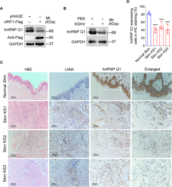
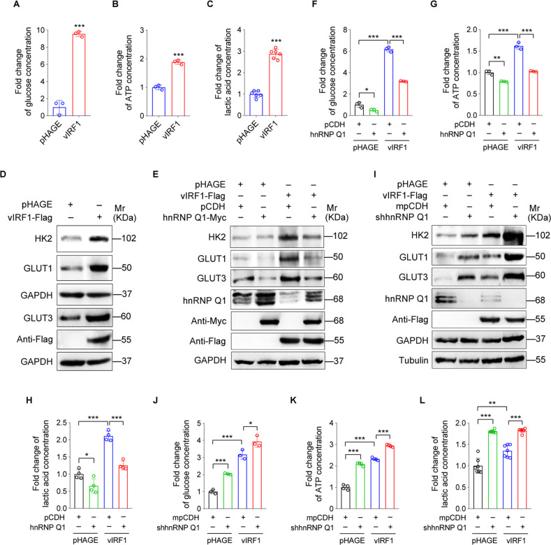
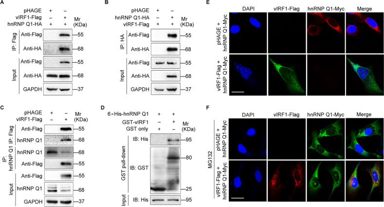
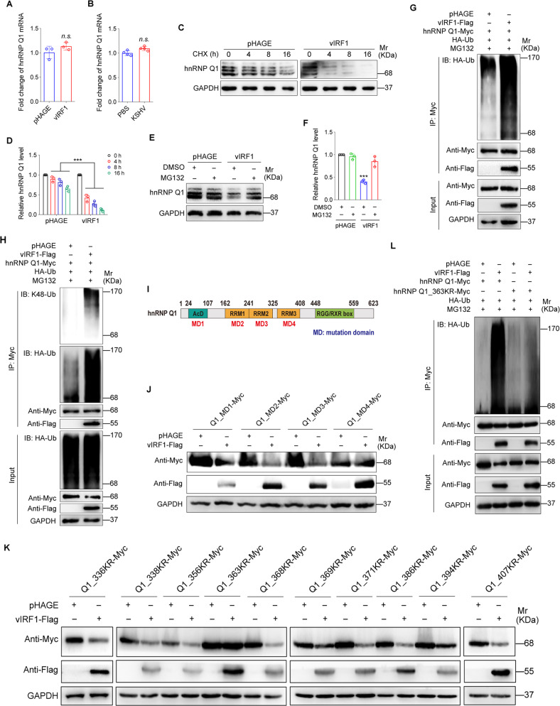
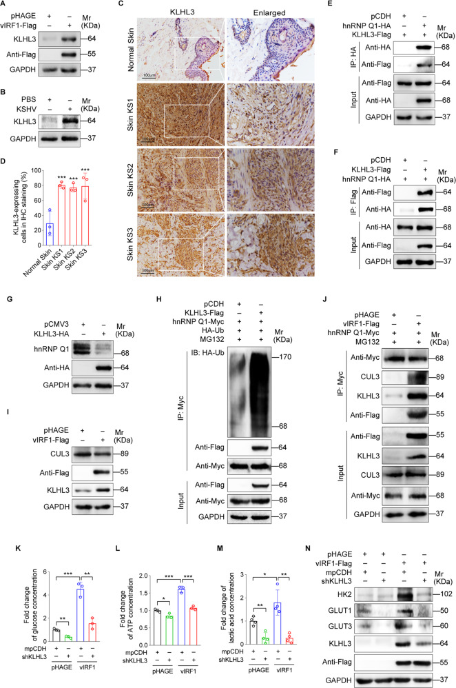
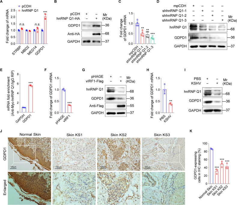
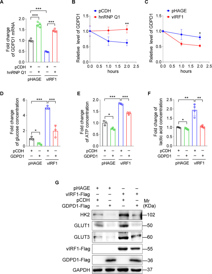
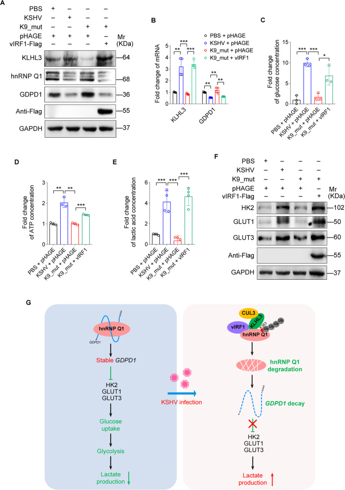
Reprogramming of host metabolism is a common strategy of viral evasion of host cells, and is essential for successful viral infection and induction of cancer in the context cancer viruses. Kaposi's sarcoma (KS) is the most common AIDS-associated cancer caused by KS-associated herpesvirus (KSHV) infection. KSHV-encoded viral interferon regulatory factor 1 (vIRF1) regulates multiple signaling pathways and plays an important role in KSHV infection and oncogenesis. However, the role of vIRF1 in KSHV-induced metabolic reprogramming remains elusive. Here we show that vIRF1 increases glucose uptake, ATP production and lactate secretion by downregulating heterogeneous nuclear ribonuclear protein Q1 (hnRNP Q1). Mechanistically, vIRF1 upregulates and recruits E3 ubiquitin ligase Kelch-like 3 (KLHL3) to degrade hnRNP Q1 through a ubiquitin-proteasome pathway. Furthermore, hnRNP Q1 binds to and stabilizes the mRNA of glycerophosphodiester phosphodiesterase domain containing 1 (GDPD1). However, vIRF1 targets hnRNP Q1 for degradation, which destabilizes GDPD1 mRNA, resulting in induction of aerobic glycolysis. These results reveal a novel role of vIRF1 in KSHV metabolic reprogramming, and identifying a potential therapeutic target for KSHV infection and KSHV-induced cancers.


Conflict of interest statement

The authors declare no competing interests.

Figures

Fig. 1
Fig. 1. vIRF1 downregulates hnRNP Q1.
A A human umbilical vein endothelial cell line EA.hy926 were transduced with 2 MOI lentiviral vIRF1 (vIRF1) or control lentivirus (pHAGE). Whole-cell lysates were collected and examined by Western blotting for hnRNP Q1 expression. B Endothelial cells EA.hy926 were treated with PBS (PBS) or infected with wild type KSHV (KSHV). Whole-cell lysates were collected and examined by Western blotting for hnRNP Q1 expression. C H&E staining, immunohistochemical staining of KSHV LANA, hnRNP Q1 in normal skin, skin KS of patient #1 (Skin KS1), skin KS of patient #2 (Skin KS2), and skin KS of patient #3 (Skin KS3). Magnification, ×200, ×400. D Results were quantified in C Data were presented with mean ± SD. ***P < 0.001, Student’s t-test.
Fig. 2
Fig. 2. vIRF1 downregulates hnRNP Q1 to promote aerobic glycolysis.
A Levels of intracellular glucose of endothelial cells transduced with 2 MOI lentiviral vIRF1 (vIRF1) or control lentivirus (pHAGE). B Levels of intracellular ATP of cells treated as in (A), C. The supernatant of cells treated as in (A) was used to measure lactate production. D Western blotting analysis of HK2, GLUT1, and GLUT3 expression in cells treated as in (A). E Lentiviral vIRF1- or its control pHAGE-infected endothelial cells were transduced with lentiviral hnRNP Q1 and its control pCDH. Whole-cell lysates were harvested and examined by Western blotting for HK2, GLUT1 and GLUT3 expression. F Cells treated as in (E) were used to examine the levels of intracellular glucose. G Cells treated as in (E) were used to measure the levels of intracellular ATP. H Cells treated as in (E) were used to measure lactate production. I Lentiviral vIRF1- or its control pHAGE-infected endothelial cells were transduced with lentivirus expressing a mixture of shRNAs targeting hnRNP Q1 (shhnRNP Q1) and its control mpCDH. Whole-cell lysates were harvested and examined by Western blotting for HK2, GLUT1 and GLUT3 expression. J Cells treated as in (I) were used to examine the levels of intracellular glucose. K Cells treated as in I were used to measure the levels of intracellular ATP. (L). Cells treated as in (I) were used to measure lactate production. Data were presented with mean ± SD. *P < 0.05, **P < 0.01, and ***P < 0.001, Student’s t-test.
Fig. 3
Fig. 3. vIRF1 directly interacts with hnRNP Q1.
A Endothelial cells transfected with hnRNP Q1-HA construct were transduced with lentiviral vIRF1-Flag or its control pHAGE. The interaction between vIRF1 and hnRNP Q1 proteins was examined by immunoprecipitating with anti-Flag antibody. B Endothelial cells transfected with vIRF1-Flag construct were transduced with lentiviral hnRNP Q1-HA or its control pHAGE. The interaction between vIRF1 and hnRNP Q1 proteins was examined by immunoprecipitating with anti-HA antibody. C Lentiviral vIRF1- or its control pHAGE-infected endothelial cells were used to examine the interaction between vIRF1 and hnRNP Q1 proteins by immunoprecipitating with anti-Flag antibody or anti-hnRNP Q1 antibody. D GST or GST- vIRF1 fusion protein was used to pull down purified 6 xHis-hnRNP Q1 protein. E Endothelial cells transfected with vIRF1-Flag and hnRNP Q1-Myc were employed to detect the expression of vIRF1 and hnRNP Q1 by immunofluorescence staining. F Endothelial cells transfected with vIRF1-Flag and hnRNP Q1-Myc were further treated with MG132 (20 μM) for 6 h and then were employed to examine the colocalization of vIRF1 and hnRNP Q1 by immunofluorescence staining.
Fig. 4
Fig. 4. vIRF1 induces hnRNP Q1 degradation.
A qPCR analysis of hnRNP Q1 mRNA expression in endothelial cells transduced with 2 MOI lentiviral vIRF1 (vIRF1) or control lentivirus (pHAGE). B qPCR analysis of hnRNP Q1 mRNA expression in endothelial cells treated with PBS (PBS) or infected with wild-type KSHV (KSHV). C Western blotting analysis of hnRNP Q1 expression in vIRF1-expressing endothelial cells treated with CHX (20 μg/ml) for 0 h, 4 h, 8 h and 16 h. D Results were quantified in (C). E Western blotting analysis of hnRNP Q1 level in vIRF1-expressing endothelial cells treated with MG132 (20 μM) for 6 h (F) Results were quantified in (E). G Endothelial cells treated as in (A) were transfected with the HA-Ub and hnRNP Q1-Myc construct, and then treated with MG132 (20 μM) for 6 h. Immunoprecipitation assay was used for detection of hnRNP Q1 ubiquitination with anti-Myc antibody. H Cells treated as in (A) were treated with MG132 (20 μM) for 6 h. Immunoprecipitation assay was used to detect the level of K48-ubiquitinated hnRNP Q1 with anti-Myc antibody. I Schematic representation of hnRNP Q1 with the indicated locations of the AcD and RRM globular domains and the RGG/RXR box motif. J Generation of the mutation constructs of hnRNP Q1 in which Lys was replaced with Arg (K to R) based on four domains of hnRNP Q1. Lentiviral vIRF1 or its control pHAGE transduced cells were transfected with plasmids encoding Myc-tagged wild type hnRNP Q1 or its mutation domain constructs (MD1, MD2, MD3 and MD4), respectively. Western blotting was performed to examine hnRNP Q1 expression with anti-Myc antibody. K Generation of the mutation constructs of hnRNP Q1 in which Lys was replaced with Arg (K to R) based on mutation domain 4 of hnRNP Q1. Lentiviral vIRF1 or its control pHAGE transduced cells were transfected with plasmids encoding 9 mutation constructs in mutation domain 4 (MD4) of Myc-tagged hnRNP Q1 (336KR, 338KR, 356KR, 363KR, 368KR, 369KR, 371KR, 386KR, 394KR and 407KR), respectively. Western blotting was performed to detect hnRNP Q1 expression level with anti-Myc antibody. L Cells treated as in (A) were transfected with the HA-Ub and hnRNP Q1-Myc or hnRNP Q1_363KR-Myc construct, and then treated with MG132 (20 μM) for 6 h. Cells were subjected to immunoprecipitation assay (IP) for detection of hnRNP Q1 ubiquitination with anti-Myc antibody. Data were presented with mean ± SD. ***P < 0.001, Student’s t-test. n.s., not significant.
Fig. 5
Fig. 5. vIRF1 recruits the E3 ubiquitin ligase KLHL3 to degrade hnRNP Q1 and promote aerobic glycolysis.
A Western blotting analysis of KLHL3 expression in endothelial cells transduced with 2 MOI lentiviral vIRF1 (vIRF1) or control lentivirus (pHAGE). B Western blotting analysis of KLHL3 expression in endothelial cells treated with PBS (PBS) or infected with wild type KSHV (KSHV). C H&E staining, and immunohistochemical staining of KLHL3 in normal skin, skin KS of patient #1 (Skin KS1), skin KS of patient #2 (Skin KS2), and skin KS of patient #3 (Skin KS3). Magnification, ×200, ×400. D Results were quantified in C. E KLHL3 overexpressed endothelial cells were transduced with lentiviral HA-tagged hnRNP Q1 or its control virus pCDH. The interaction between KLHL3 and hnRNP Q1 proteins was examined by immunoprecipitating with anti-HA antibody. F hnRNP Q1 overexpressed endothelial cells were transduced with lentiviral Flag-tagged KLHL3 or its control virus pCDH. The interaction between KLHL3 and hnRNP Q1 proteins was examined by immunoprecipitating with anti-Flag antibody. G Western blotting analysis of hnRNP Q1 expression in endothelial cells which were transfected with pCMV3-Flag-KLHL3 construct (KLHL3) or its control pCMV3-C-Flag (pCMV). H The HA-Ub and hnRNP Q1-Myc plasmids were co-transfected into endothelial cells. Cells were further transduced with lentiviral KLHL3-Flag or its control virus pCDH, and treated with MG132 (20 μM) for 6 h. Cells were subjected to immunoprecipitation assay (IP) with anti-Myc antibody for detection of hnRNP Q1 ubiquitination. I Western blotting analysis of CUL3 and KLHL3 expression in endothelial cells transduced with 2 MOI lentiviral vIRF1 (vIRF1) or control lentivirus (pHAGE). J hnRNP Q1 (hnRNP Q1-Myc) overexpressed endothelial cells were transduced with lentiviral vIRF1 and its control lentivirus pHAGE, and then further treated with MG132 (20 μM) for 6 h. Immunoprecipitation assay was performed to examine the interaction between hnRNP Q1 and KLHL3 /CUL3 complex with anti-Myc antibody. K Lentiviral vIRF1- or its control pHAGE-infected endothelial cells, followed by transduction with lentivirus expressing a mixture of shRNAs targeting KLHL3 (shKLHL3) were used to detect the levels of intracellular glucose. L Cells treated as in (K) were used to measure the levels of intracellular ATP. M. Cells treated as in (K) were used to measure lactate production. N Western blotting analysis of HK2, GLUT1 and GLUT3 expression in cells treated as in (K). Data were shown as mean ± SD. * P < 0.05, ** P < 0.01, and *** P < 0.001, Student’s t-test.
Fig. 6
Fig. 6. GDPD1 mRNA is the target of hnRNP Q1.
A qRT-PCR analysis of STRBP, MBD2, MED14 and GDPD1 mRNA levels in endothelial cells transduced with lentiviral hnRNP Q1 (hnRNP Q1) and its control lentivirus (pCDH). B Western blotting analysis of GDPD1 protein level in cells treated as in (A). C qRT-PCR analysis of GDPD1 mRNA level in endothelial cells transduced with lentivirus-mediated No.1 (shhnRNP Q1-1), No.2 (shhnRNP Q1-2) and No. 3 (shhnRNP Q1-3) shRNAs targeting hnRNP Q1. D Western blotting analysis of GDPD1 expression in cells treated as in (C). E RNA immunoprecipitation analysis of the interaction between hnRNP Q1 and GDPD1 mRNA with anti-Myc antibody. GAPDH was used as a control. F qRT-PCR analysis of GDPD1 mRNA level in endothelial cells transduced with 2 MOI lentiviral vIRF1 (vIRF1) or control lentivirus (pHAGE). G Western blotting analysis of GDPD1 expression in endothelial cells transduced with 2 MOI lentiviral vIRF1 (vIRF1) or control lentivirus (pHAGE). H qRT-PCR analysis of GDPD1 mRNA level in endothelial cells treated with PBS (PBS) or infected with wild type KSHV (KSHV). I Western blotting analysis of GDPD1 expression in endothelial cells treated with PBS (PBS) or infected with wild type KSHV (KSHV). J H&E staining, and immunohistochemical staining of GDPD1 protein in normal skin, skin KS of patient #1 (Skin KS1), skin KS of patient #2 (Skin KS2), and skin KS of patient #3 (Skin KS3). Magnification, ×200, ×400. K Results were quantified in J Data were shown as mean ± SD. **P < 0.01, and ***P < 0.001, Student’s t-test. n.s., not significant.
Fig. 7
Fig. 7. vIRF1-downregulated hnRNP Q1 reduces the stability of GDPD1 mRNA to induce aerobic glycolysis.
A Lentiviral vIRF1- or its control pHAGE-infected endothelial cells were transduced with lentiviral hnRNP Q1 and its control lentivirus pCDH. Cells were collected for qRT-PCR analysis of GDPD1 mRNA level. B Endothelial cells transduced with lentiviral hnRNP Q1 or control lentivirus pCDH were treated with actinomycin D. RNA decay assays were performed to examine the degradation rate of GDPD1 mRNA. C Endothelial cells transduced with 2 MOI lentiviral vIRF1 (vIRF1) or control lentivirus (pHAGE) were treated with actinomycin D. RNA decay assays were performed to detect the degradation rate of GDPD1 mRNA. D Lentiviral vIRF1- or its control pHAGE-infected endothelial cells were transduced by lentiviral GDPD1 (GDPD1-Flag) and control lentivirus (pCDH). Cells were used to detect the level of intracellular glucose. E Cells treated as in (D) were used to measure the levels of intracellular ATP. F Cells treated as in (D) were used to measure lactate production. G Western blotting analysis of HK2, GLUT1 and GLUT3 expression in cells treated as in (D). Data were shown as mean ± SD. *P < 0.05, **P < 0.01, and ***P < 0.001, Student’s t-test.
Fig. 8
Fig. 8. Loss of vIRF1 reduces KSHV-induced aerobic glycolysis.
A Western blotting analysis of KLHL3, hnRNP Q1 and GDPD1 expression in endothelial cells treated with PBS (PBS), infected with wild-type KSHV (KSHV) or K9 mutant virus (K9_mut) followed by transduction with lentiviral vIRF1. B qRT-PCR analysis of KLHL3 and GDPD1 mRNA levels in cells treated as in (A). C Cells treated as in (A) were used to examine the level of intracellular glucose. D Cells treated as in (A) were used to measure the level of intracellular ATP. E Cells treated as in (A) were used to detect lactate production. F Western blotting analysis of HK2, GLUT1 and GLUT3 expression in cells treated as in (A). G Schematic illustration for the mechanism of vIRF1-induced aerobic glycolysis. KSHV vIRF1 recruits E3 ubiquitin ligase complex KLHL3/CUL3 to degrade RNA-binding protein hnRNP Q1. The downregulated hnRNP Q1 accelerates GDPD1 mRNA decay to increase glucose uptake, and lactate secretion. Data were shown as mean ± SD. *P < 0.05, **P < 0.01, and ***P < 0.001, Student’s t-test.

Similar articles

Cited by

References

    1. Bannai H, Fukatsu K, Mizutani A, Natsume T, Iemura S, Ikegami T, et al. An RNA-interacting protein, SYNCRIP (heterogeneous nuclear ribonuclear protein Q1/NSAP1) is a component of mRNA granule transported with inositol 1,4,5-trisphosphate receptor type 1 mRNA in neuronal dendrites. J Biol Chem. 2004;279:53427–34. doi: 10.1074/jbc.M409732200. - DOI - PubMed
    1. Cesarman E, Damania B, Krown SE, Martin J, Bower M, Whitby D. Kaposi sarcoma. Nat Rev Dis Prim. 2019;5:9. doi: 10.1038/s41572-019-0060-9. - DOI - PMC - PubMed
    1. Bhutani M, Polizzotto MN, Uldrick TS, Yarchoan R. Kaposi sarcoma-associated herpesvirus-associated malignancies: epidemiology, pathogenesis, and advances in treatment. Semin Oncol. 2015;42:223–46. doi: 10.1053/j.seminoncol.2014.12.027. - DOI - PMC - PubMed
    1. Soulier J, Grollet L, Oksenhendler E, Cacoub P, Cazals-Hatem D, Babinet P, et al. Kaposi’s sarcoma-associated herpesvirus-like DNA sequences in multicentric Castleman’s disease. Blood. 1995;86:1276–80. doi: 10.1182/blood.V86.4.1276.bloodjournal8641276. - DOI - PubMed
    1. Warburg O, Wind F, Negelein E. The metabolism of tumors in the body. J Gen Physiol. 1927;8:519–30. doi: 10.1085/jgp.8.6.519. - DOI - PMC - PubMed

Publication types

MeSH terms

Substances