Review
. 2022 Nov;12(11):2613-2633.
doi: 10.1007/s13346-022-01138-1. Epub 2022 May 10.

Transdermal delivery for gene therapy

Affiliations
Review

Transdermal delivery for gene therapy

Parbeen Singh et al. Drug Deliv Transl Res. 2022 Nov.

Abstract

Gene therapy is a critical constituent of treatment approaches for genetic diseases and has gained tremendous attention. Treating and preventing diseases at the genetic level using genetic materials such as DNA or RNAs could be a new avenue in medicine. However, delivering genes is always a challenge as these molecules are sensitive to various enzymes inside the body, often produce systemic toxicity, and suffer from off-targeting problems. In this regard, transdermal delivery has emerged as an appealing approach to enable a high efficiency and low toxicity of genetic medicines. This review systematically summarizes outstanding transdermal gene delivery methods for applications in skin cancer treatment, vaccination, wound healing, and other therapies.

Keywords: DNA; Gene therapy; Microneedles; Transdermal delivery; mRNA; miRNA; siRNA.


Conflict of interest statement

TDN has a conflict of interest with PiezoBioMembrane Inc. and SingleTimeMicroneedles Inc.

Figures

Fig. 1
Fig. 1
Schematic of different transdermal gene therapy methods
Fig. 2
Fig. 2
Schematic of transdermal microneedle patch delivery (A) and intracellular pathway (B) of genome editing agent (Cas9) nanocomplex and glucocorticoid nanoparticle (dexamethasone delivered from the patch) to treat the inflammatory skin diseases. T7 endonuclease I assay of indels (method to validate CRISPR reagents) introduced into the nod-like receptor family, pyrin domain-containing 3 (NLRP3) locus of DC (dendritic cell) 2.4 cells (C) and 3T3 cells (D) transfected with the two nanoparticles. The figure was reproduced from ref. [38]. Copyright Science Advance 2021
Fig. 3
Fig. 3
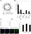
Schematic of SPACE-peptide decorated cationic ethosomes for transdermal siRNA delivery (A). Skin permeability of peptide system under various conditions (B). Internalization of model drug by SPACE peptide after 30 min (C) and 120 min (D) incubation time. FITC internalization after 120-min incubation with various concentrations (E). Cell toxicity of SPACE peptide with 1 mg (open bar) and 10 mg (close bar) with keratinocytes for different time periods (F). The figure was reproduced from ref. [65]. Copyright Elsevier 2014
Fig. 4
Fig. 4
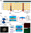
Ultrasound-based transdermal therapies. Schematic of multi-transdermal techniques (MNs, ultrasound, and iontophoresis) for transdermal drug delivery (A-B). Dissolution mechanism by ultrasound (C) and predicted dissolution mechanism and ion direction (D). The figure was reproduced from ref. [83]. Copyright Nature publishing group 2020
Fig. 5
Fig. 5
Schematic of iontophoresis-based anti-STAT3 siRNA and imatinib mesylate delivery (A). STAT3 protein expression in mice after treatment (B). Quantified tumor weights after treatment with different formulations (C) and change in volume of tumor (D). The figure was reproduced from ref. [95]. Copyright Elsevier 2017
Fig. 6
Fig. 6
Schematic model of the skin–electrode interface, SC and its iconic pathways (A). The figure was reproduced from ref. [102]. Copyright Plos One 2015. Schematic of electroporation device with distinct layers (B). Digital image of electroporation device sandwiched between two ITO electrodes (C). Schematic of concept of localize electroporation and asymmetric FEM stimulation of electric field along with single nano-channel underneath a cell (D). Pore evaluation model (left) and molecular transport model (right) (E). Electroporation-based delivery of PI into HT 1080 cells using a 10-V pulse (F). Delivery efficiency and viability of live and dead staining after 6-h electroporation. The figure was reproduced from ref. [111]. Copyright ACS publication 2019
Fig. 7
Fig. 7
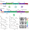
Schematic of prokaryotic and eukaryotic mRNA (A). The figure was adopted from Ref. [127]. Copyright Elsevier 2016. Transfection efficiency and expression of 5 μg luciferase mRNA via MNs with different heights:H400 (B), H800 (C), and H1000 (D) in the mice with different time points. Fluorescence images after 6-h transfection (E). Luciferase expression in the mice by MNs and subcutaneous injection (F). The figure was reproduced from ref. [146]. Copyright Nature publication 2018
Fig. 8
Fig. 8
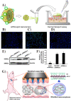
Schematic of FTIC attached lipid nanocarriers for transdermal delivery (A). Comparison of DOTAP-NC and lipofectamine effect on GAPDH knockdown in HeLa cells after 6 h, control; only free siRNA (B), positive control; lipo-siRNA (C); DOTAP-NC-siRNA (D). GAPDH gene expression using by western blotting (E) and relative gene expression (F). The figure was reproduced from ref. [175]. Copyright Wiley–VCH 2020. Schematic of R8/siBARF coated MNs for tumor therapy (G). The figure was reproduced from ref. [180]. Copyright Elsevier 2018
Fig. 9
Fig. 9
Transdermal miRNA-based gene therapy. Schematic of miRNA-21 mimic BA-PEI nano-carrier for wound healing (A) and effect of miRNA-21 in wound healing (B). The figure was reproduced from [191]. Copyright Theranostic publication. MNs functionalized with be spoke peptide nucleic acid probe with covalently bound to alginate hydrogel via photo-cleavable linker (C). Protocol for MN-based sampling of target biomarker and purification to remove the non-target sequence (D). The figure was reproduced from ref. [192]. Copyright ACS publications 2019. Schematic of transdermal delivery of pDNA encoding miRNA-221 inhibitor gene by NPs for skin cancer treatment (E–H). The figure was reproduced from ref. [27]. Copyright ACS publication 2017. The miRNA-1331 delivered by ultrasound with low frequency suppressed the tumor growth and improved the survival rate. Tumor images after therapy (I), tumor growth during therapy (J), Survival rate with scramble-miRNA-MB (K), and survival rate with low frequency ultrasound (L). The figure was reproduced from ref. [193]. Copyright Wiley VCH 2016
Fig. 10
Fig. 10
Schematic of MN-based DNA delivery to prevent the cervical cancer (A). The figure was reproduced from ref. [203]. Copy right Elsevier 2017. Fluorescence images of cell EGFP gene expression in COS-7 cells transfected with DNA release from coated MNs over 0–3 h (B-C) and 36–47 h using Lipofectamine 2000. The figure was reproduced from ref. [204]. Copyright ACS publication. Average tumor volume of control and immunized mice (D). The figure was reproduced from ref. [205]. Copyright Elsevier 2019

Similar articles

Cited by

References

    1. Dunbar CE, High KA, Joung JK, Kohn DB, Ozawa K, Sadelain M. Gene therapy comes of age. Science. 2018;359(6372). 10.1126/science.aan4672. - PubMed
    1. Bulaklak K, Gersbach CA. The once and future gene therapy. Nat Commun. 2020;11(1):5820. doi: 10.1038/s41467-020-19505-2. - DOI - PMC - PubMed
    1. Pipe SW. Gene therapy for hemophilia. Pediatr Blood Cancer. 2018;65(2):e26865. doi: 10.1002/pbc.26865. - DOI - PubMed
    1. Wolf DP, Mitalipov PA, Mitalipov SM. Principles of and strategies for germline gene therapy. Nat Med. 2019;25(6):890–897. doi: 10.1038/s41591-019-0473-8. - DOI - PubMed
    1. FDA. What is gene therapy. 2019 a.

LinkOut - more resources