Evaluation of root canal morphology in permanent maxillary and mandibular anterior teeth in Saudi subpopulation using two classification systems: a CBCT study
- PMID: 35538514
- PMCID: PMC9092761
- DOI: 10.1186/s12903-022-02187-1
Evaluation of root canal morphology in permanent maxillary and mandibular anterior teeth in Saudi subpopulation using two classification systems: a CBCT study
Abstract
Background: Adequate knowledge of root canal morphology and possible variations is essential to achieve perfect root canal treatment and overcome treatment failure. Appropriate knowledge on root and canal morphology, communication, and documentation amongst dentists will be challenging from a diagnostic and successful treatment point of view.
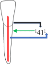
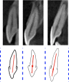
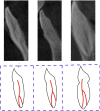
Methods: A total of 3420 samples were included in this study from 285 cone-beam computed tomography images of the Saudi residents, including 171 males and 114 females aged 15 to 68 years from retrospective data dated from January 2018 to April 2021. The images were examined in sagittal, axial and coronal views using a 3D version software 1.0.10.6388. The number of canal and canal morphology was recorded using Vertucci and the new classification system. The SPSS 26 was used to conduct the statistical analysis as descriptive statistics such as mean; standard deviation and frequency were calculated. The Chi-square test analysed the data with the significance level set at 0.05.
Results: A total of 285 subjects participated in the study. Majority of the participants were Saudi nationals (80.7%), followed by Indian (7.4%), Pakistani (4.2%) and other nationalities. According to Vertucci and the new classification system, Type I and 1TN1 were the most common types, followed by Type III and Type IV, and then 1TN1-2-1 and 1TN1-2 in mandibular anteriors. The prevalence of canal variations in mandibular canine was higher in females than in males (P = 0.002). Maxillary laterals and mandibular anteriors showed the significant difference in the prevalence of root canal variation in relation to the ethnicity (P = 0.001) and age of the patients. Younger patients showed more variations than the older patients (P = 0.012, P = 0.023, P = 0.001, P = 0.001) in terms of maxillary laterals, mandibular central, laterals and canines, respectively.
Conclusion: Mandibular permanent anteriors showed a wide range of canal variations and canal complexity. Males and females did not demonstrate a wide range of variation in the root canal morphology except for the canines in relation to the gender of the patients.
Keywords: Classification; Dental Pulp; Dental anatomy; Endodontics; Morphology; Root canal.
© 2022. The Author(s).
Conflict of interest statement
The authors declare no conflict of interest.
Figures


Similar articles
-
Evaluation of root and canal morphology of mandibular premolar amongst Saudi subpopulation using the new system of classification: a CBCT study.BMC Oral Health. 2023 May 15;23(1):291. doi: 10.1186/s12903-023-03002-1. BMC Oral Health. 2023. PMID: 37189077 Free PMC article.
-
Investigating root and canal morphology of anterior and premolar teeth using CBCT with a novel coding classification system in Saudi subpopulation.Sci Rep. 2025 Feb 5;15(1):4392. doi: 10.1038/s41598-025-86277-4. Sci Rep. 2025. PMID: 39910098 Free PMC article.
-
Root canal morphology of anterior permanent teeth in Jordanian population using two classification systems: a cone-beam computed tomography study.BMC Oral Health. 2024 Feb 2;24(1):170. doi: 10.1186/s12903-024-03934-2. BMC Oral Health. 2024. PMID: 38308267 Free PMC article.
-
Assessment of the root and canal morphology in the permanent dentition of Saudi Arabian population using cone beam computed and micro-computed tomography - a systematic review.BMC Oral Health. 2024 Mar 16;24(1):343. doi: 10.1186/s12903-024-04101-3. BMC Oral Health. 2024. PMID: 38493123 Free PMC article.
-
Evaluation of root canal morphology of human primary molars by using CBCT and comprehensive review of the literature.Acta Odontol Scand. 2016;74(4):250-8. doi: 10.3109/00016357.2015.1104721. Epub 2015 Nov 2. Acta Odontol Scand. 2016. PMID: 26523502 Review.
Cited by
-
Bibliometric analysis: Root and root canal morphology using cone-beam computed tomography.Clin Exp Dent Res. 2023 Dec;9(6):1156-1168. doi: 10.1002/cre2.801. Epub 2023 Oct 25. Clin Exp Dent Res. 2023. PMID: 37877522 Free PMC article. Review.
-
Evaluation of root and canal morphology of mandibular premolar amongst Saudi subpopulation using the new system of classification: a CBCT study.BMC Oral Health. 2023 May 15;23(1):291. doi: 10.1186/s12903-023-03002-1. BMC Oral Health. 2023. PMID: 37189077 Free PMC article.
-
Classifying the internal anatomy of anterior teeth in the Yemeni population using two systems: a retrospective CBCT study.Odontology. 2025 Jan;113(1):416-431. doi: 10.1007/s10266-024-00965-7. Epub 2024 Jun 27. Odontology. 2025. PMID: 38935196
-
Prevalence of Isthmi and Root Canal Configurations in Mandibular Permanent Teeth Using Cone-Beam Computed Tomography.Biomed Res Int. 2024 Jul 31;2024:9969860. doi: 10.1155/2024/9969860. eCollection 2024. Biomed Res Int. 2024. PMID: 39118804 Free PMC article.
-
Endodontic Management of a Maxillary Central Incisor with two Roots: A Case Report and Literature Review.Iran Endod J. 2023;18(3):174-180. doi: 10.22037/iej.v18i3.41296. Iran Endod J. 2023. PMID: 37431523 Free PMC article.
References
-
- Karobari MI, et al. Root and canal morphology of the anterior permanent dentition in Malaysian population using two classification systems: a CBCT clinical study. Australian Endodont J. 2020. - PubMed
Publication types
MeSH terms
LinkOut - more resources
Full Text Sources
Miscellaneous

