. 2022 May 10;29(1):31.
doi: 10.1186/s12929-022-00814-1.

Inhibition of ChREBP ubiquitination via the ROS/Akt-dependent downregulation of Smurf2 contributes to lysophosphatidic acid-induced fibrosis in renal mesangial cells

Affiliations

Inhibition of ChREBP ubiquitination via the ROS/Akt-dependent downregulation of Smurf2 contributes to lysophosphatidic acid-induced fibrosis in renal mesangial cells

Donghee Kim et al. J Biomed Sci. .

Abstract

Background: Mesangial cell fibrosis, a typical symptom of diabetic nephropathy (DN), is a major contributor to glomerulosclerosis. We previously reported that the pharmacological blockade of lysophosphatidic acid (LPA) signaling improves DN. Although LPA signaling is implicated in diabetic renal fibrosis, the underlying molecular mechanisms remain unclear. Here, the role of carbohydrate-responsive element-binding protein (ChREBP) in LPA-induced renal fibrosis and the underlying mechanisms were investigated.

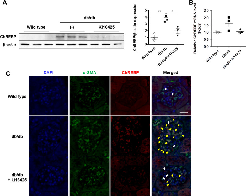
Methods: Eight-week-old wild-type and db/db mice were intraperitoneally injected with the vehicle or an LPAR1/3 antagonist, ki16425 (10 mg/kg), for 8 weeks on a daily basis, following which the mice were sacrificed and renal protein expression was analyzed. SV40 MES13 cells were treated with LPA in the presence or absence of ki16425, and the expression of ChREBP and fibrotic factors, including fibronectin, TGF-β, and IL-1β, was examined. The role of ChREBP in the LPA-induced fibrotic response was investigated by ChREBP overexpression or knockdown. The involvement of Smad ubiquitination regulatory factor-2 (Smurf2), an E3 ligase, in LPA-induced expression of ChREBP and fibrotic factors was investigated by Smurf2 overexpression or knockdown. To identify signaling molecules regulating Smurf2 expression by LPA, pharmacological inhibitors such as A6370 (Akt1/2 kinase inhibitor) and Ly 294002 (PI3K inhibitor) were used.

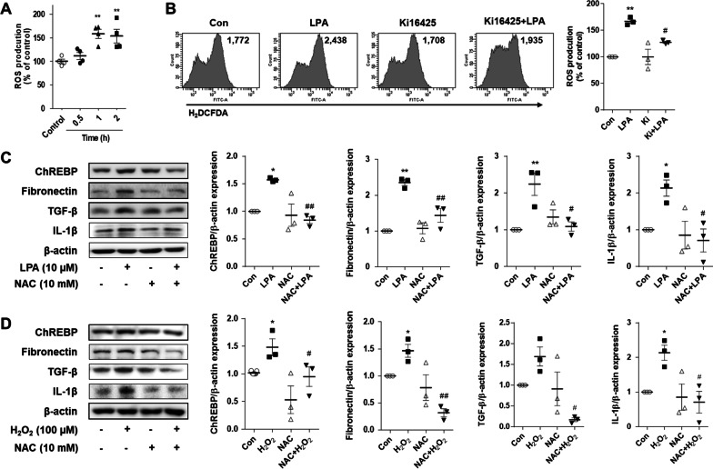
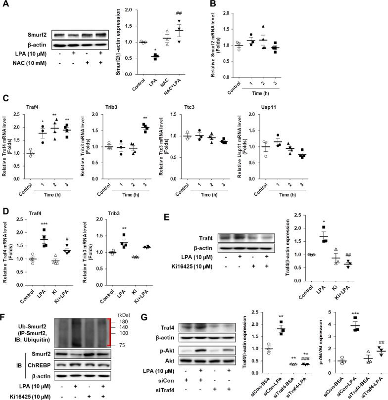
Results: The renal expression of ChREBP increased in diabetic db/db mice, and was reduced following treatment with the ki16425. Treatment with LPA induced the expression of ChREBP and fibrotic factors, including fibronectin, TGF-β, and IL-1β, in SV40 MES13 cells, which were positively correlated. The LPA-induced expression of fibrotic factors increased or decreased following ChREBP overexpression and knockdown, respectively. The production of reactive oxygen species (ROS) mediated the LPA-induced expression of ChREBP and fibrotic factors, and LPA decreased Smurf2 expression via Traf4-mediated ubiquitination. The LPA-induced expression of ubiquitinated-ChREBP increased or decreased following Smurf2 overexpression and knockdown, respectively. Additionally, Smurf2 knockdown significantly increased the expression of ChREBP and fibrotic factors. The pharmacological inhibition of Akt signaling suppressed the LPA-induced alterations in the expression of ChREBP and Smurf2.

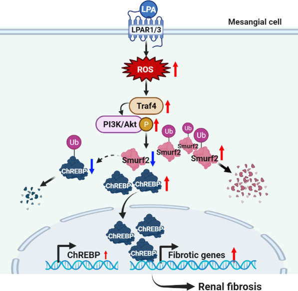
Conclusion: Collectively, the results demonstrated that the ROS/Akt-dependent downregulation of Smurf2 and the subsequent increase in ChREBP expression might be one of the mechanisms by which LPA induces mesangial cell fibrosis in DN.

Keywords: ChREBP; Diabetic nephropathy; Fibrosis; Lysophosphatidic acid; Mesangial cells; Smurf2.


Conflict of interest statement

All the authors declared no competing interest.

Figures

Fig. 1
Fig. 1
ChREBP expression is reduced in the renal cortex of db/db mice treated with the LPAR inhibitor, ki16425. Eight-week-old wild-type and db/db mice were intraperitoneally injected with the vehicle or ki16425 (10 mg/kg) for 8 weeks on a daily basis, following which the mice were sacrificed. A The level of ChREBP protein in the renal cortex was analyzed by western blotting, quantified using ImageJ software, and normalized to those of β-actin. B The level of ChREBP mRNA in the renal cortex was determined by quantitative real-time polymerase chain reaction (qRT-PCR). The data are presented as the mean ± SEM (n = 3/group); *p < 0.05, **p < 0.01. C Representative images depicting the colocalization of α-SMA (green) and ChREBP (red) in the kidney tissue sections of mice. The nuclei were counterstained with DAPI (blue). The white arrows indicate the cytosolic expression of ChREBP, and the yellow arrows indicate the nuclear expression of ChREBP in α-SMA-positive cells. Scale bars, 20 μm; n = 3
Fig. 2
Fig. 2
LPA induces the expression of ChREBP and fibrotic factors in SV40 MES13 cells. A SV40 MES13 cells were treated with 10 µM LPA for the indicated durations. The level of ChREBP protein was analyzed by western blotting, quantified using ImageJ software, and normalized to that of β-actin. The data are presented as the mean ± SEM of results obtained from three independent experiments. *p < 0.05 vs. control. B SV40 MES13 cells were seeded in 4-well chambers and treated with LPA in the presence or absence of ki16425 for 3 h. Immunocytochemistry analysis was performed using an anti-ChREBP antibody and an Alexa Fluor 546-conjugated secondary antibody. The nuclei were counterstained with DAPI (blue) (original magnification, 400 ×; the scale bars represent 20 μm). C The SV40 MES13 cells were treated with LPA in the presence or absence of ki16425 for 3 h. The protein levels of ChREBP, fibronectin, TGF-β, and IL-1β were analyzed by western blotting, quantified using ImageJ software, and normalized to those of β-actin. The data are presented as the mean ± SEM of results obtained from three independent experiments. *p < 0.05 vs. control (Con); #p < 0.05 vs. LPA
Fig. 3
Fig. 3
ChREBP regulates the LPA-induced expression of fibrotic factors in SV40 MES13 cells. A and B SV40 MES13 cells were transfected with a control siRNA (siCon) or ChREBP siRNA (siChREBP), and treated with 10 µM LPA for 3 h. The mRNA (A) and protein (B) levels of ChREBP, fibronectin, TGF-β, and IL-1β were analyzed by qRT-PCR (n = 5) and western blotting (n = 3), respectively. C and D The SV40 MES13 cells were co-transfected with vectors expressing ChREBP and Mlx (ChREBP/Mlx), or transfected with an empty pcDNA vector (Con), and treated with 10 µM LPA for 3 h. The mRNA (C) and protein (D) levels of ChREBP, fibronectin, TGF-β, and IL-1β were analyzed by qRT-PCR (n = 5) and western blotting (n = 3), respectively. The data are presented as the mean ± SEM. *p < 0.05, **p < 0.01, ***p < 0.005 vs. siCon-BSA or Con-BSA; #p < 0.05, ##p < 0.01, ###p < 0.005 vs. siCon-LPA or Con-LPA
Fig. 4
Fig. 4
LPA induces the production of ROS, and ROS scavenging decreases the LPA- or ROS-induced expression of ChREBP and fibrotic factors in SV40 MES13 cells. A SV40 MES13 cells were treated with 10 µM LPA for the indicated durations. B SV40 MES13 cells were treated with LPA in the presence or absence of ki16425 for 1 h. A and B The intracellular levels of ROS were measured by flow cytometry with DCFH-DA. The values in the representative flow cytometry histograms indicate the fluorescence intensity of DCF in the whole cells. The relative fluorescence intensity levels of DCF were compared with the production of ROS in the control cells. C The cells were pretreated with NAC for 1 h and subsequently treated with LPA for 3 h. D The cells were pretreated with NAC for 1 h and subsequently treated with H2O2 for 1 h. C and D The protein levels of ChREBP, fibronectin, TGF-β, and IL-1β were analyzed by western blotting, quantified using ImageJ software, and normalized to those of β-actin. The data are presented as the mean ± SEM of results obtained from three independent experiments. *p < 0.05, **p < 0.01 vs. Con; #p < 0.05, ##p < 0.01 vs. LPA or H2O2
Fig. 5
Fig. 5
LPA induces the expression of ChREBP and fibrotic factors via Smurf2 in SV40 MES13 cells. A SV40 MES13 cells were treated with LPA, either in the presence or absence of ki16425, or treated with Heclin (Hec) for 3 h. B and C The SV40 MES13 cells were transfected with a control siRNA (siCon) or Smurf2 siRNA (siSmurf2, siSm2) for 6 h, then the medium was replaced with SFM for 16–18 h, and treated with LPA for 3 h. The proteins levels of ChREBP, Smurf2, fibronectin, TGF-β, and IL-1β were analyzed by western blotting, quantified using ImageJ software, and normalized to those of β-actin. The data are presented as the mean ± SEM of results obtained from three to five independent experiments. *p < 0.05, **p < 0.01 vs. Con or siCon-BSA; #p < 0.05, ##p < 0.01, ###p < 0.005 vs. LPA or siCon-LPA
Fig. 6
Fig. 6
LPA inhibits the ubiquitination of ChREBP by downregulating Smurf2 in SV40 MES13 cells. A SV40 MES13 cells were treated with LPA in the presence or absence of ki16425 for 3 h. B The cells were transfected with a control siRNA (siCon) or Smurf2 siRNA (siSmurf2) for 6 h, then the medium was replaced with SFM for 16–18 h, and subsequently treated with LPA for 3 h. C The cells were transfected with pCMV5B-Flag-Smurf2 wt (Smurf2 wt) or pCMV5B-Flag-Smurf2 C716A (Smurf2 C716A) for 6 h, then the medium was replaced with SFM for 16–18 h, and subsequently treated with LPA for 3 h. (A-C) The cell lysates were subjected to immunoprecipitation studies by incubating with an anti-ChREBP antibody followed by blotting with anti-ubiquitin antibody. The protein levels of ubiquitinated-ChREBP (Ub-ChREBP), ChREBP, and Smurf2 were analyzed by western blotting. Representative images of the blots from three independent experiments are depicted, where the red brackets indicate Ub-ChREBP
Fig. 7
Fig. 7
Inhibition of Akt signaling suppresses the alterations in the expression of ChREBP and Smurf2 induced by LPA. A SV40 MES13 cells were pretreated with A6730 (Akt1/2 kinase inhibitor, 4 μM) or Ly 294002 (PI3K inhibitor, 10 μM) for 1 h, and subsequently treated with LPA and/or ki16425 for 3 h. B SV40 MES13 cells were pretreated with NAC for 1 h and subsequently treated with LPA for 3 h. A and B The protein levels of p-Akt, ChREBP, and Smurf2 were analyzed by western blotting, quantified using ImageJ software, and normalized to those of β-actin. The data are presented as the mean ± SEM of results obtained from four independent experiments. *p < 0.05, **p < 0.01 vs. vehicle only (control); #p < 0.05, ##p < 0.01 vs. LPA only
Fig. 8
Fig. 8
LPA increases Traf4-mediated ubiquitination of Smurf2 in SV40 MES13 cells. A SV40 MES13 cells were pretreated with NAC for 1 h and subsequently treated with LPA for 3 h. The level of Smurf2 protein was analyzed by western blotting, quantified using ImageJ software, and normalized to that of β-actin. B and C SV40 MES13 cells were treated with 10 µM LPA for the indicated durations. The mRNA levels of Smurf2 (B) and ubiquitin ligases (C), including Traf4, Trib3, Ttc3, and USP11, were determined by qRT-PCR. DF SV40 MES13 cells were treated with LPA in the presence or absence of ki16425 for 3 h. D The mRNA levels of Traf4 and Trib3 were determined by qRT-PCR. E The level of Traf4 protein was analyzed by western blotting, quantified using ImageJ software, and normalized to that of β-actin. F The cell lysates were subjected to immunoprecipitation studies by incubating with an anti-Smurf2 antibody followed by blotting with anti-ubiquitin antibody. The protein levels of ubiquitinated-Smurf2 (Ub-Smurf2), ChREBP, and Smurf2 were analyzed by western blotting. Representative images of the blots from three independent experiments are depicted, where the red brackets indicate Ub-Smurf2. G SV40 MES13 cells were transfected with a control (siCon) or Traf4 (siTraf4) siRNA for 6 h, then the medium was replaced with SFM for 16–18 h, and treated with LPA for 3 h. The proteins levels of Traf4 and p-Akt were analyzed by western blotting, quantified using ImageJ software, and normalized to those of β-actin and Akt, respectively. The data are presented as the mean ± SEM of results obtained from four independent experiments. *p < 0.05, **p < 0.01 vs. vehicle only (control) or siCon-BSA; #p < 0.05, ##p < 0.01 vs. LPA only or siCon-LPA
Fig. 9
Fig. 9
Smurf2 expression is restored in the glomeruli of db/db mice treated with the ki16425. Eight-week-old wild-type and db/db mice were intraperitoneally injected with the vehicle or ki16425 (10 mg/kg/day) for 8 weeks and the mice were sacrificed. Immunohistochemical detection of Smurf2 was performed in the kidney tissues using DAB. The nuclei were counterstained with hematoxylin. The representative images are depicted (original magnification, 200 ×; scale bars, 20 μm; n = 3)
Fig. 10
Fig. 10
Schematic representation of the mechanism by which LPA induces ChREBP-mediated fibrosis, via the ROS/Akt-dependent downregulation of Smurf2 in renal mesangial cells. Created with BioRender.com. Red and blue arrows indicate upregulated and downregulated responses, respectively

Similar articles

Cited by

References

    1. Bhattacharjee N, Barma S, Konwar N, Dewanjee S, Manna P. Mechanistic insight of diabetic nephropathy and its pharmacotherapeutic targets: an update. Eur J Pharmacol. 2016;791:8–24. doi: 10.1016/j.ejphar.2016.08.022. - DOI - PubMed
    1. Alsaad KO, Herzenberg AM. Distinguishing diabetic nephropathy from other causes of glomerulosclerosis: an update. J Clin Pathol. 2007;60(1):18–26. doi: 10.1136/jcp.2005.035592. - DOI - PMC - PubMed
    1. Lim A. Diabetic nephropathy—complications and treatment. Int J Nephrol Renov Dis. 2014;7:361–381. doi: 10.2147/IJNRD.S40172. - DOI - PMC - PubMed
    1. Zhao JH. Mesangial cells and renal fibrosis. Adv Exp Med Biol. 2019;1165:165–194. doi: 10.1007/978-981-13-8871-2_9. - DOI - PubMed
    1. Lin ME, Herr DR, Chun J. Lysophosphatidic acid (LPA) receptors: signaling properties and disease relevance. Prostaglandins Other Lipid Mediat. 2010;91(3–4):130–138. doi: 10.1016/j.prostaglandins.2009.02.002. - DOI - PMC - PubMed

MeSH terms

Substances