Review
. 2022 May 10;20(1):206.
doi: 10.1186/s12967-022-03405-8.

Patient-derived xenograft (PDX) models, applications and challenges in cancer research

Affiliations
Review

Patient-derived xenograft (PDX) models, applications and challenges in cancer research

Shahrokh Abdolahi et al. J Transl Med. .

Abstract

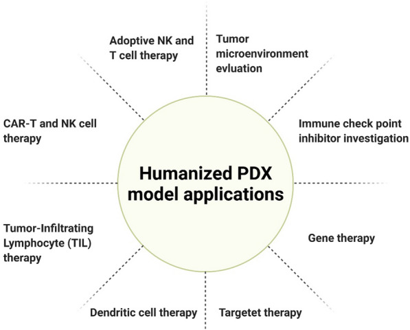
The establishing of the first cancer models created a new perspective on the identification and evaluation of new anti-cancer therapies in preclinical studies. Patient-derived xenograft models are created by tumor tissue engraftment. These models accurately represent the biology and heterogeneity of different cancers and recapitulate tumor microenvironment. These features have made it a reliable model along with the development of humanized models. Therefore, they are used in many studies, such as the development of anti-cancer drugs, co-clinical trials, personalized medicine, immunotherapy, and PDX biobanks. This review summarizes patient-derived xenograft models development procedures, drug development applications in various cancers, challenges and limitations.

Keywords: Avatar model of cancer; Cancer animal model; Humanized model; PDX; Preclinical study.


Conflict of interest statement

The authors declare that they have no competing interests.

Figures

Fig. 1
Fig. 1
Cancer animal models timeline. The timeline shows the first available reports of the use of animal models during the Aristotle and Erasistratus eras, which over time, recognizing how cancer formed in 1777 and identifying carcinogens paved the way for the use of cancerous animal models in 1910 with the development of DBA mice. Finally, in 1918, the first cancer model was developed by Yamagiwa and Ichikawa. Following the development of the first cell line in 1951, the first CDX model was introduced, which greatly contributed to the improvement of cancer science. Furthermore, PDX and GEMMs were introduced in 1969 and 1981, respectively. GEMMs genetically engineered mouse models, PDX patient-derived xenograft, CDX Cell Line-Derived Xenograft model, DBA dilute, brown and non-agouti
Fig. 2
Fig. 2
Cancer animal models over the years. The animal model of cancer was first introduced in 1918, but the beginning of today’s models was in 1951 with the development of the CDX models, but the development of other models led to competition for a more suitable model with more efficiency. The beginning of the twenty-first century can be considered the beginning of the flourishing of animal models of cancer. CDX model won the competition between the CDX and GEMME models because of its availability and is still the first choice in many studies. But the remarkable thing is that at the beginning of the second decade of the twenty-first century, PDX models have attracted attention and are being used in various studies with considerable speed. Data obtained from the PubMed database. GEMMs genetically engineered mouse models, PDX patient-derived xenograft, CDX Cell Line-Derived Xenograft model)
Fig. 3
Fig. 3
The process of creating PDX models and their applications. The generations are named F1, F2, F3, etc. Different cancer tissues can be used to create PDX models (orthotopic or heterotopic engraftment), as well as different hosts with varying degrees of immune deficiency. Tumor biopsy and generations F1 and F2 can be sampled for tissue bank, and tumor biopsy and F3 can be used for Genomics, Proteomics, and Transcriptomics analyzes. The F3 generation of these models can be used in various studies. CTC circulating tumor cell, S.C subcutaneously, I.V intravenous, I.P intraperitoneal, FFPE formalin-fixed paraffin-embedded
Fig. 4
Fig. 4
PDX clinical trial and Co-clinical trial. A In models that follow the PDX clinical trial approach, a large number of PDXs originate from several patients or samples of a bio-repository. Each model tests a specific drug regimen, and the information obtained is evaluated. B Co-clinical trials were developed to achieve precision medicine. In fact, the mouse trials are performed in parallel with the human trials, and then in real-time, the information obtained from the mouse study is transferred to the human study and integrated. This leads to the most effective clinical outcome
Fig. 5
Fig. 5
Humanized PDX model applications in immunotherapy

References

    1. Pott P. Chirurgical observations relative to the cataract, the polypus of the nose, the cancer of the scrotum, the different kinds of ruptures, and the mortification of the toes and feet, by Percivall Pott, FRS Surgeon to St. Bartholomew’s-Hospital. Printed, by TJ Carnegy, For L. Hawes, W. Clarke, and R. Collins, in Pater-noster Row; 1775.
    1. Hedrich HJ. The house mouse as a laboratory model: a historical perspective. The Laboratory Mouse. Burlington: Elsevier Science; 2012. p. 868.
    1. Johnson J, et al. Relationships between drug activity in NCI preclinical in vitro and in vivo models and early clinical trials. Br J Cancer. 2001;84(10):1424–1431. doi: 10.1054/bjoc.2001.1796. - DOI - PMC - PubMed
    1. Ellis LM, Fidler IJ. Finding the tumor copycat: therapy fails, patients don’t. Nat Med. 2010;16(9):974–975. doi: 10.1038/nm0910-974. - DOI - PubMed
    1. Rezaee R, Abdollahi M. The importance of translatability in drug discovery. Expert Opin Drug Discov. 2017;12(3):237–239. doi: 10.1080/17460441.2017.1281245. - DOI - PubMed