. 2018 May 23;8(34):18972-18979.
doi: 10.1039/c8ra02804d. eCollection 2018 May 22.

Cyclization of secondarily structured oligonucleotides to single-stranded rings by using Taq DNA ligase at high temperatures

Affiliations

Cyclization of secondarily structured oligonucleotides to single-stranded rings by using Taq DNA ligase at high temperatures

Yixiao Cui et al. RSC Adv. .

Abstract

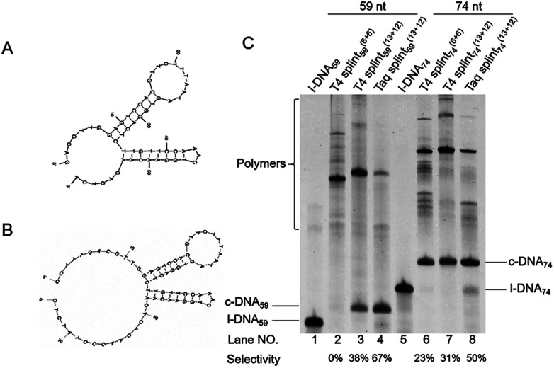
Single-stranded DNA rings play important roles in nanoarchitectures, molecular machines, DNA detection, etc. Although T4 DNA ligase has been widely employed to cyclize single-stranded oligonucleotides into rings, the cyclization efficiency is very low when the oligonucleotides (l-DNAs) take complicated secondary structures at ambient temperatures. In the present study, this problem has been solved by using Thermus aquaticus DNA ligase (Taq DNA ligase) at higher temperatures (65 and 70 °C) where the secondary structures are less stable or completely destroyed. This method is based on our new finding that this ligase successfully functions even when the splint strand is short and forms no stable duplex with l-DNA (at least in the absence of the enzyme). In order to increase the efficiency of cyclization, various operation factors (lengths and sequences of splint, as well as the size of the DNA ring) have been investigated. Based on these results, DNA rings have been successfully synthesized from secondarily structured oligonucleotides in high yields and high selectivity. The present methodology is applicable to the preparation of versatile DNA rings involving complicated secondary structures, which should show novel properties and greatly widen the scope of DNA-based nanotechnology.


Conflict of interest statement

None declared.

Figures

Scheme 1
Scheme 1. Schematic view of cyclization of single-stranded linear DNA (l-DNA) by DNA ligase. In pathway 1, l-DNA takes no secondary structure at 25 °C and is successfully cyclized by T4 DNA ligase with the assistance of splint DNA. In pathway 2, however, l-DNA is folded to a complicated secondary structure at 25 °C, and thus is hardly cyclized by T4 DNA ligase at 25 °C, even in the presence of splint DNA. In pathway 3, this problem is solved by using Taq DNA ligase and achieving the reactions at higher temperatures where the secondary structures are unstable. The ring structure of the final product is confirmed in terms of the resistance against the digestion by Exonuclease I.
Fig. 1
Fig. 1. Cyclization of l-DNAs by Taq DNA ligase and T4 DNA ligase. Both l-DNA59 (A) and l-DNA74 (B) take secondary structures, as determined by Mfold calculation ([Mg2+] = 10 mM at 25 °C). In (C), the cyclization was carried out under the following conditions: [l-DNA]0 = 5 μM and [12 or 25 nt splint] = 10 μM. Lane 1, l-DNA59 only; lane 2, DNA59 with splint59(6+6) at 25 °C by T4 DNA ligase; lane 3, l-DNA59 with splint59(13+12) at 25 °C by T4 DNA ligase; lane 4, l-DNA59 with splint59(13+12) at 65 °C by Taq DNA ligase; lane 5, l-DNA74 only; lane 6, l-DNA74 with splint74(6+6) at 25 °C by T4 DNA ligase; lane 7, l-DNA74 with splint74(13+12) at 25 °C by T4 DNA ligase; lane 8, l-DNA74 with splint74(13+12) at 65 °C by Taq DNA ligase. Prior to the reactions, l-DNAs were phosphorylated at the 5′-position by using T4 polynucleotide kinase. The sequences of l-DNAs and splints are listed in Table 1 and detailed reaction conditions are shown in Experimental section. The Tm of l-DNA59 and l-DNA74 was 65.1 °C and 66.4 °C (Fig. S1†).
Fig. 2
Fig. 2. Effects of the length of splint on the cyclization of l-DNA74 by Taq DNA ligase at (A) 65 °C and (B) 70 °C. The reaction conditions: [l-DNA74]0 = 5 μM; [splint] = 10 μM; 40 U Taq DNA ligase (in 20 μL). In (C) and (D), the yields and the selectivity at 65 °C (circles) and 70 °C (triangles) are plotted as a function of splint length. The results in lanes 2 and 3 in (A) and lanes 2–4 in (B) were not incorporated to the plots in (D), since the band intensities of the products were too small to determine their ratios precisely. The sequences of splints are listed in Table S1.
Fig. 3
Fig. 3. Cyclization of l-DNA (74 nt) by Taq DNA ligase using the splints of various GC contents. Reaction conditions: [l-DNA]0 = 5 μM, [splint] = 10 μM, and 40 U Taq DNA ligase (in 20 μL) at 65 °C for 12 h. The sequences of all the l-DNAs and the splints are shown in Table S1.
Fig. 4
Fig. 4. Schematic of view of cyclization of l-DNA of different lengths by Taq DNA ligase (A). The reaction conditions in (B): [l-DNA64, l-DNA54, or l-DNA44]0 = 5 μM, [splint74(15+14)] = 10 μM, and 40 U Taq DNA ligase (in 20 μL) at 65 °C for 12 h. For the purpose of comparison, [splint74(6+6)] = 10 μM and the results with T4 DNA ligase at 25 °C are also presented. The sequences of l-DNAs and splints are listed in Table S1.
Fig. 5
Fig. 5. Diluted buffer improves the selectivity. (A) Effect of the concentration of Taq DNA ligase buffer on the cyclization of l-DNA74 by Taq DNA ligase. Diluted buffers were employed under the conditions: [l-DNA74]0 = 5 μM, [splint74(15+14)] = 10 μM, and 40 U Taq DNA ligase (in 20 μL) for 12 h. (B) Effect of the initial concentration of l-DNA74 in 0.1× Taq DNA ligase buffer at 70 °C. [l-DNA74]0/[splint74(15+14)] = 1/2. For the purpose of comparison, the results with T4 DNA ligase in 0.1× T4 DNA ligase buffer are also shown. In lanes 6 and 7 in (B), the products in lanes 2 and 4, respectively, were treated with Exonuclease I to digest non-cyclic DNAs.
Scheme 2
Scheme 2. Species formed in the mixtures for the cyclization of oligonucleotide by Taq DNA ligase.

Similar articles

Cited by

References

    1. Wang K. He M. Q. Zhai F. H. Wang J. He R. H. Yu Y. L. Biosens. Bioelectron. 2018;105:159–165. doi: 10.1016/j.bios.2018.01.044. - DOI - PubMed
    1. Xu H. Jiang Y. Liu D. Liu K. Zhang Y. Yu S. Shen Z. Wu Z. S. Anal. Chim. Acta. 2018;1011:86–93. doi: 10.1016/j.aca.2018.01.022. - DOI - PubMed
    1. Teller C. Willner I. Curr. Opin. Biotechnol. 2010;21:376–391. doi: 10.1016/j.copbio.2010.06.001. - DOI - PubMed
    1. Komiyama M. Yoshimoto K. Sisido M. Ariga K. Bull. Chem. Soc. Jpn. 2017;90:967–1004. doi: 10.1246/bcsj.20170156. - DOI
    1. Gaillard C. Strauss F. PLoS One. 2015;10:e0119368. - PMC - PubMed