. 2018 Jul 16;8(45):25480-25488.
doi: 10.1039/c8ra03943g.

Preparation of porous calcium phosphate microspheres with phosphate-containing molecules at room temperature for drug delivery and osteogenic differentiation

Affiliations

Preparation of porous calcium phosphate microspheres with phosphate-containing molecules at room temperature for drug delivery and osteogenic differentiation

Jun-Feng Liu et al. RSC Adv. .

Abstract

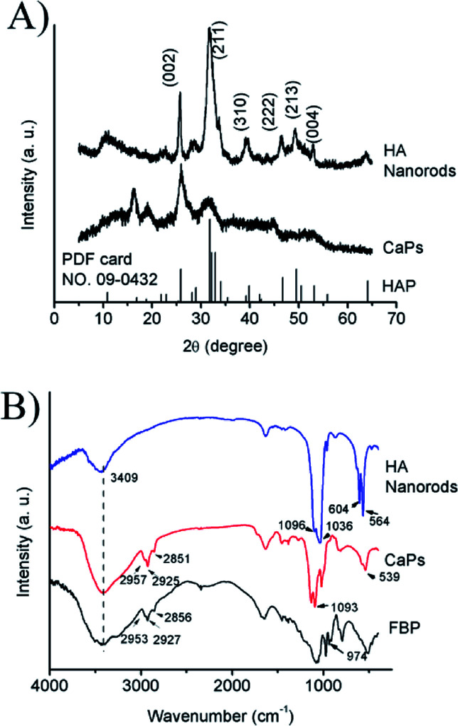
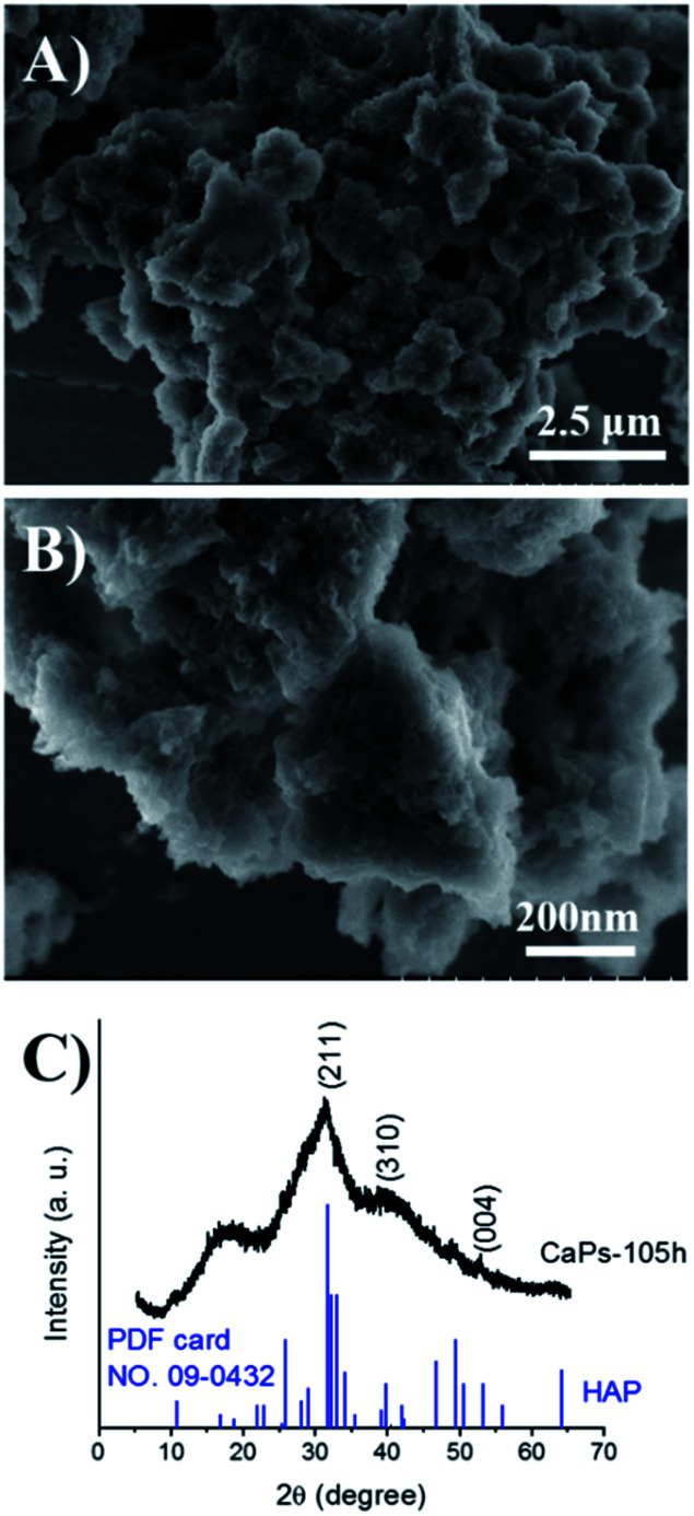
Calcium phosphate (CaP) has similar chemical properties to those of the inorganic component of human bone tissue, for potential application in drug delivery for the chemotherapy of osteosarcoma. In this work, CaP with a porous microsphere structure has been synthesized using fructose-1,6-bisphosphate (FBP) as the phosphorus source by a simple wet-chemical strategy at room temperature. The CaP porous microspheres, as an organic-inorganic hybrid nano-platform, exhibit good doxorubicin (Dox) loading capacity, and Dox-loading CaP, enhancing the in vitro chemotherapy of osteosarcoma cells. The CaP porous microspheres show high biocompatibility, and induce the osteogenic differentiation of MC3T3-E1. These results indicate that the CaP porous microspheres reported in this study are promising for application as an anti-osteosarcoma drug carrier and osteoinductive material for bone regeneration in the treatment of osteosarcoma.


Conflict of interest statement

There are no conflicts to declare.

Figures

Fig. 1
Fig. 1. SEM and TEM micrographs of HA samples (A and B) and CaP porous microspheres (C–F). The inset in B shows the SAED pattern of the HA samples, and the inset in F shows the SAED pattern of the CaP porous microspheres.
Fig. 2
Fig. 2. (A) PXRD patterns of the CaP porous microspheres and HA samples; (B) FTIR spectra of pure FBP, the CaP porous microspheres and HA nanorods.
Scheme 1
Scheme 1. Schematic illustration of the strategy for the preparation of CaP porous microspheres using FBP disodium salt as a phosphorus source.
Fig. 3
Fig. 3. The N2 adsorption–desorption isotherm of CaP porous microspheres synthesized using FBP molecules as the phosphorus source.
Fig. 4
Fig. 4. Dox release curves of the Dox-loaded CaPs and Dox-loaded HA in PBS solutions (pH 7.4, 37 °C).
Fig. 5
Fig. 5. SEM micrographs (A and B) and PXRD pattern (C) of the CaP porous microspheres after being immersed in PBS solution (pH 7.4) at 37 °C for 105 h.
Fig. 6
Fig. 6. Cell viability studies of the HA nanorods and CaP porous microspheres. (A) Cell viability assay of MC3T3-E1 cells co-cultured with CaP porous microspheres or HA nanorods for 1 day. (B) Cell viability assay of MC3T3-E1 cells co-cultured with the extract solution of CaP porous microspheres or HA nanorods at different concentrations for 1, 3 and 7 days.
Fig. 7
Fig. 7. Dox-loaded CaP porous microspheres show a better inhibitory effect on cell proliferation than Dox-loaded HA nanorods on MG-63 cells (A) and 143b cells (B) co-cultured for 1 day at various concentrations. The inhibition rate of the MG-63 cells (C) and 143b cells (D) co-cultured with Dox-loaded CaP porous microspheres (CaPs-Dox) and Dox-loaded HA nanorods (HA-Dox) for two days at various concentrations.
Fig. 8
Fig. 8. Increased Dox fluorescence intensity in MG-63 cells after being co-cultured with Dox-loaded HA nanorods (HA-Dox) and Dox-loaded CaP porous microspheres (CaPs-Dox) for 24 h.
Fig. 9
Fig. 9. DAPI staining and phalloidine staining of MG-63 cells when co-cultured with Dox-loaded HA nanorods (HA-Dox) and Dox-loaded CaP porous microspheres (CaPs-Dox) for 24 h.
Fig. 10
Fig. 10. 143b cells were co-cultured with CaP porous microspheres (CaPs-Dox) and HA-Dox for one day at a concentration of 5 μg mL−1.
Fig. 11
Fig. 11. The CaP porous microspheres induced higher ALP activity than the HA nanorods at certain concentrations and co-culture times in the MC3T3-E1 cells. *Significant difference (p < 0.05) compared with the HA nanorods at the same concentration.
Fig. 12
Fig. 12. von Kossa staining (A) and alizarin red staining (B) of the calcium nodules of MC3T3-E1 cells co cultured with the CaP porous microspheres and HA nanorods.
Fig. 13
Fig. 13. Schematic illustration of the functions of CaP porous microspheres in osteosarcoma treatment. CaP porous microspheres display dual functions, to deliver Dox to inhibit the proliferation of osteosarcoma cells and promote the osteogenic differentiation of MC3T3-E1.

Similar articles

Cited by

References

    1. Harrison D. J. Geller D. S. Gill J. D. Lewis V. O. Gorlick R. Expert Rev. Anticancer Ther. 2018;18:39–50. doi: 10.1080/14737140.2018.1413939. - DOI - PubMed
    1. Lindsey B. A. Markel J. E. Kleinerman E. S. Rheumatol Ther. 2017;4:25–43. doi: 10.1007/s40744-016-0050-2. - DOI - PMC - PubMed
    1. Bielack S. S. Hecker-Nolting S. Blattmann C. Kager L. F1000Research. 2016;5:2767. - PMC - PubMed
    1. Zhou Z. F. Sun T. W. Chen F. Zuo D. Q. Wang H. S. Hua Y. Q. Cai Z. D. Tan J. Biomaterials. 2017;121:1–14. doi: 10.1016/j.biomaterials.2016.12.031. - DOI - PubMed
    1. Son K. D. Kim Y. J. Biomater. Res. 2017;21:13. doi: 10.1186/s40824-017-0099-1. - DOI - PMC - PubMed