. 2018 Feb 23;8(16):8455-8468.
doi: 10.1039/c8ra00296g.

Retracted Article: Linc00472 suppresses breast cancer progression and enhances doxorubicin sensitivity through regulation of miR-141 and programmed cell death 4

Affiliations

Retracted Article: Linc00472 suppresses breast cancer progression and enhances doxorubicin sensitivity through regulation of miR-141 and programmed cell death 4

Pengwei Lu et al. RSC Adv. .

Retraction in

Abstract

The existence of drug resistance strikingly hampers the therapy of many malignancies, including breast cancer. Long non-coding RNAs (LncRNAs) have been reported to participate in the regulation of various biological processes associated with cancer progression. Whereas, the role of linc00472 in breast cancer pathogenesis and doxorubicin (ADR) resistance have not been well elucidated. In the present study, it is found that linc00472 expression was decreased in breast cancer tissues and cells. Moreover, higher linc00472 expression was positively associated with favorable disease status and prognosis for breast cancer patients. Functional analyses revealed that linc00472 overexpression suppressed proliferation and invasion, facilitated apoptosis and enhanced ADR sensitivity in breast cancer cells. Mechanistic studies discovered that linc00472 acted as a competing endogenous RNA (ceRNA) of miR-141 to sequester miR-141 from its target mRNA PDCD4 (programmed cell death 4). Furthermore, the inhibition effect of linc00472 on breast cancer cell progression and ADR resistance could be partly abrogated by miR-141 up-regulation or PDCD4 knockdown. In vivo assays also demonstrated that linc00472 hindered tumor growth by suppressing miR-141 expression and enhancing PDCD4 expression. In conclusion, linc00472 blocked breast cancer progression and induced ADR sensitivity through regulation of miR-141 and PDCD4, highlighting a potential therapeutic strategy for breast cancer patients.


Conflict of interest statement

The authors have no conflict of interest to declare.

Figures

Fig. 1
Fig. 1. Linc00472 expression was down-regulated in breast cancer tissues and cells. RT-qPCR assays were carried out to measure the expression patterns of linc00472 in 40 pairs of breast cancer tissues and adjacent normal tissues (A), in breast cancer tissues with different degree of lymph-node metastasis (N0, N1, N2, N3) (B), in breast cancer tissues with (n = 15) or without (n = 25) distant metastasis (C), in human normal mammary epithelial cell line MCF-10A and three breast cancer cell lines (MDA-MB-231, MDA-MB-453, MCF-7) (D). (E) Kaplan–Meier curve was employed to assess the overall survival outcome in breast cancer patients with high-level linc00472 expression (n = 17) and low-level linc00472 expression (n = 15). *P < 0.05.
Fig. 2
Fig. 2. Linc00472 overexpression suppressed breast cancer cells proliferation and invasion. MCF-7 and MDA-MB-453 cells were transfected with pcDNA3.1 or pcDNA-linc00472 vector. (A) RT-qPCR assay was performed to measure linc00472 expression at 48 h after transfection. (B and C) MTT assay was used to evaluate cell viability at the indicated time points (0, 24, 48, 72 h) posttransfection. (D and E) EdU-positive cells percentage was detected by EdU immunofluorescence staining and flow cytometric analysis at 48 h after transfection. (F) Transfected cells were seeded in 6-well plates and incubated for 15 days, then cell colony number was assessed. (G and H) Cell cycle distribution was determined by flow cytometry 48 h posttransfection. (I and J) Cell invasion capability was monitored using transwell invasion assays at 48 h after transfection. *P < 0.05.
Fig. 3
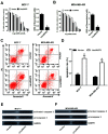
Fig. 3. Linc00472 overexpression increased doxorubicin (ADR) chemosensitivity and induced ADR-mediated apoptosis in breast cancer cells. (A and B) MCF-7 cells and MDA-MB-453 cells were transfected with pcDNA3.1 vector or pcDNA-linc00472 for 24 h, followed by treatment with different concentration of ADR (0.001, 0.01, 0.1, 1, 10, 100 μM) for 48 h. Then MTT assays were performed to determine cell survival rate and IC50 values of ADR. (C–F) MCF-7 cells and MDA-MB-453 cells transfected with pcDNA3.1 vector or pcDNA-linc00472 were treated with 0.1 μM ADR for 48 h, followed by analysis of apoptotic rate by flow cytometry (C and D), as well as assessment of activated caspase 3 and procaspase 3 protein levels by western blot (E and F). *P < 0.05.
Fig. 4
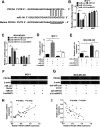
Fig. 4. Linc00472 directly inhibited miR-141 expression. (A) The predicted binding sites between linc00472 and miR-141 together with the mutant sites in the linc00472-MUT vector. (B and C) RNA of nucleus and cytoplasm in MCF-7 and MDA-MB-453 cells was isolated by subcellular fractionation assays. Then expressions of GAPDH, U6 snRNA and linc00472 were quantified by RT-qPCR assays with U6 snRNA as a nucleus control and GAPDH as a cytoplasm control. (D and E) MCF-7 cells were co-transfected with linc00472-WT or linc00472-MUT reporter and miR-con or miR-141, and MDA-MB-453 cells were co-transfected with linc00472-WT or linc00472-MUT reporter and anti-miR-con or anti-miR-141. At 48 h after transfection, luciferase activity was detected by Dual-Luciferase reporter assays. (F and G) RIP assays were performed using IgG or Ago2 antibody in MCF-7 and MDA-MB-453 cells, followed by the detection of linc00472 and miR-141 expression in immunoprecipitate. IgG performed as a negative control and input as a positive control. (H and I) RT-qPCR assay was carried out to analyze the expressions linc00472 and miR-141 in MCF-7 and MDA-MB-453 cells transfected with si-linc00472 or si-con. (J) miR-141 expression was detected in MCF-7 and MDA-MB-453 cells transfected with pcDNA3.1, pcDNA-linc00472 or pcDNA-linc00472-MUT vector using RT-qPCR assay. (K) Spearman's correlation analysis of linc00472 and miR-141 expression in 40 cases of breast cancer tissues. *P < 0.05.
Fig. 5
Fig. 5. Linc00472 up-regulated PDCD4 expression by sponging miR-141. (A) The putative binding sites between PDCD4 and miR-141 by TargetScan website and the mutant sites in the mutant-PDCD4 luciferase reporter vector. (B) MCF-7 cells were co-transfected with wide-type or mutant-type PDCD4-3′UTR reporter and miR-con, miR-141, miR-141+pcDNA3.1 vector, or miR-141+pcDNA-linc00472, followed by the detection of luciferase activity 48 h posttransfection. (C) MDA-MB-453 cells were co-transfected with wide-type or mutant-type PDCD4-3′UTR reporter and anti-miR-con, anti-miR-141, anti-miR-141+si-con, or anti-miR-141+si-linc00472, followed by the analysis of luciferase activity at 48 h after transfection. (D–G) MCF-7 cells were transfected with miR-con+vector, miR-141+vector, miR-con+linc00472, or miR-141+linc00472, and MDA-MB-453 cells were transfected with anti-miR-con+si-con, anti-miR-141+si-con, anti-miR-con+si-linc00472, or anti-miR-141+si-linc00472. At 48 h after transfection, miR-141 expression level was detected by RT-qPCR assays (D and E) and PDCD4 protein level was measured using western blot assays (F and G). (H and I) Spearman's correlation analysis between linc00472 or miR-141 and PDCD4 in 40 cases of breast cancer tumor tissues. *P < 0.05.
Fig. 6
Fig. 6. miR-141 overexpression or PDCD4 knockdown abolished the inhibition effect of linc00472 on breast cancer cell progression. MCF-7 cells were transfected with vector+miR-con, linc00472+miR-con, vector + miR-141 or linc00472+miR-141, and MDA-MB-453 cells were transfected with vector+si-con, linc00472+si-con, vector+si-PDCD4 or linc00472+si-PDCD4, followed by PDCD4 protein levels detection using western blot assays (A and B), cell proliferation ability assessment by MTT assays (C and D) and colony formation assays (E), invasion capability evaluation with transwell invasion assays (F), as well as ADR IC50 values determination through MTT assays (G). *P < 0.05.
Fig. 7
Fig. 7. Linc00472 suppressed tumor growth via modulating miR-141 and PDCD4. MCF-7 cells stably transfected with lenti-con (control), lenti-pre-miR-141, lenti-linc00472 or lenti-linc00472+ lenti-pre-miR-141 were subcutaneously injected into the armpit of nude mice, followed by the detection of tumor volume at 7, 14, 21, 28, 35, 42 days after injection (A), photograph of representative tumors at 42 days after injection (B), measurement of tumor weight at 42 days after inoculation (C), assessment of miR-141 and linc00472 expression levels (D), analysis of PDCD4 and ki-67 protein expression levels (E). *P < 0.05.

References

    1. Siegel R. L. Miller K. D. Jemal A. Ca-Cancer J. Clin. 2016;66:7–30. doi: 10.3322/caac.21332. - DOI - PubMed
    1. Cardoso F. Castiglione M. Group E. G. W. Ann. Oncol. 2009;22:15–18. - PubMed
    1. Gamucci T. D'Ottavio A. M. Magnolfi E. Barduagni M. Vaccaro A. Sperduti I. Moscetti L. Belli F. Meliffi L. Br. J. Cancer. 2007;97:1040–1045. doi: 10.1038/sj.bjc.6603982. - DOI - PMC - PubMed
    1. Desantis C. E. Fedewa S. A. Goding S. A. Kramer J. L. Smith R. A. Jemal A. Ca-Cancer J. Clin. 2015;66:31–42. doi: 10.3322/caac.21320. - DOI - PubMed
    1. Sledge G. W. Neuberg D. Bernardo P. Ingle J. N. Martino S. Rowinsky E. K. Wood W. C. J. Clin. Oncol. 2003;21:588–592. doi: 10.1200/JCO.2003.08.013. - DOI - PubMed

Publication types