

Lac water extract inhibits IFN-γ signaling through JAK2-STAT1-IRF1 axis in human melanoma
- PMID: 35539920
- PMCID: PMC9080938
- DOI: 10.1039/c8ra02955e
Lac water extract inhibits IFN-γ signaling through JAK2-STAT1-IRF1 axis in human melanoma
Abstract
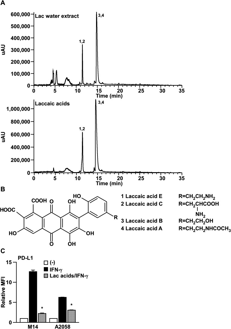
Interferon-γ (IFN-γ) is a cytokine that plays an important role in the host defense of infectious diseases and in immune surveillance during tumor development; however, it has adverse effects in the pathogenesis of autoimmune diseases and in immunosuppressive microenvironments, promoting the immunoevasion of cancer cells. In this study, we identified lac water extract (Lac) as a candidate that can suppress IFN-γ signaling amongst 112 types of natural products, using PD-L1 promoter as a readout for the IFN-γ signaling. Moreover, we determined that Lac inhibits IFN-γ-induced PD-L1 and MHC class I expression on the cell surface in melanoma cells, both of which have been identified as the downstream molecules of IFN-γ signaling. We also determined that Lac inhibited the JAK2-STAT1-IRF1 pathway. Finally, we identified laccaic acids, encompassing laccaic acid A, B, C, and E, as the active components in Lac that inhibit IFN-γ signaling. Collectively, the laccaic acids are lead compounds for a novel inhibitor that targets the JAK2-STAT1-IRF1 pathway for diseases caused by the aberrant activation of IFN-γ signaling.
This journal is © The Royal Society of Chemistry.
Conflict of interest statement
There are no conflicts of interest to declare.
Figures


Similar articles
-
PD-L1 upregulation in myeloma cells by panobinostat in combination with interferon-γ.Oncotarget. 2019 Mar 8;10(20):1903-1917. doi: 10.18632/oncotarget.26726. eCollection 2019 Mar 8. Oncotarget. 2019. PMID: 30956773 Free PMC article.
-
IFN-γ-mediated inhibition of lung cancer correlates with PD-L1 expression and is regulated by PI3K-AKT signaling.Int J Cancer. 2018 Aug 15;143(4):931-943. doi: 10.1002/ijc.31357. Epub 2018 Mar 25. Int J Cancer. 2018. PMID: 29516506
-
Genetic defects of the IRF1-mediated major histocompatibility complex class I antigen presentation pathway occur prevalently in the JAK2 gene in non-small cell lung cancer.Oncotarget. 2017 May 8;8(37):60975-60986. doi: 10.18632/oncotarget.17689. eCollection 2017 Sep 22. Oncotarget. 2017. PMID: 28977839 Free PMC article.
-
miR-375 inhibits IFN-γ-induced programmed death 1 ligand 1 surface expression in head and neck squamous cell carcinoma cells by blocking JAK2/STAT1 signaling.Oncol Rep. 2018 Mar;39(3):1461-1468. doi: 10.3892/or.2018.6177. Epub 2018 Jan 2. Oncol Rep. 2018. PMID: 29328389
-
Association of IFN-gamma signal transduction defects with impaired HLA class I antigen processing in melanoma cell lines.Clin Cancer Res. 2011 May 1;17(9):2668-78. doi: 10.1158/1078-0432.CCR-10-2114. Epub 2011 Jan 19. Clin Cancer Res. 2011. PMID: 21248298 Free PMC article.
Cited by
-
From immune checkpoints to therapies: understanding immune checkpoint regulation and the influence of natural products and traditional medicine on immune checkpoint and immunotherapy in lung cancer.Front Immunol. 2024 Feb 15;15:1340307. doi: 10.3389/fimmu.2024.1340307. eCollection 2024. Front Immunol. 2024. PMID: 38426097 Free PMC article. Review.
-
MicroRNA-721 regulates gluconeogenesis via KDM2A-mediated epigenetic modulation in diet-induced insulin resistance in C57BL/6J mice.Biol Res. 2024 May 14;57(1):27. doi: 10.1186/s40659-024-00495-0. Biol Res. 2024. PMID: 38745315 Free PMC article.
References
LinkOut - more resources
Full Text Sources
Research Materials
Miscellaneous