

Core/shell upconversion nanoparticles with intense fluorescence for detecting doxorubicin in vivo
- PMID: 35539931
- PMCID: PMC9081841
- DOI: 10.1039/c8ra02928h
Core/shell upconversion nanoparticles with intense fluorescence for detecting doxorubicin in vivo
Erratum in
-
Correction: Core/shell upconversion nanoparticles with intense fluorescence for detecting doxorubicin in vivo.RSC Adv. 2018 Jul 2;8(42):23930. doi: 10.1039/c8ra90055h. eCollection 2018 Jun 27. RSC Adv. 2018. PMID: 35544026 Free PMC article.
Abstract
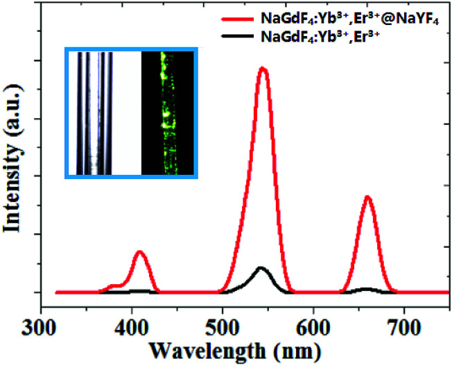
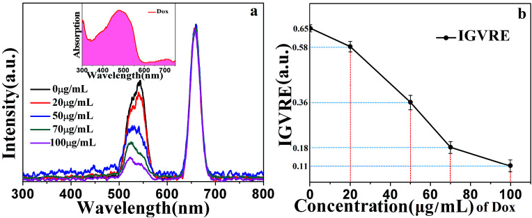
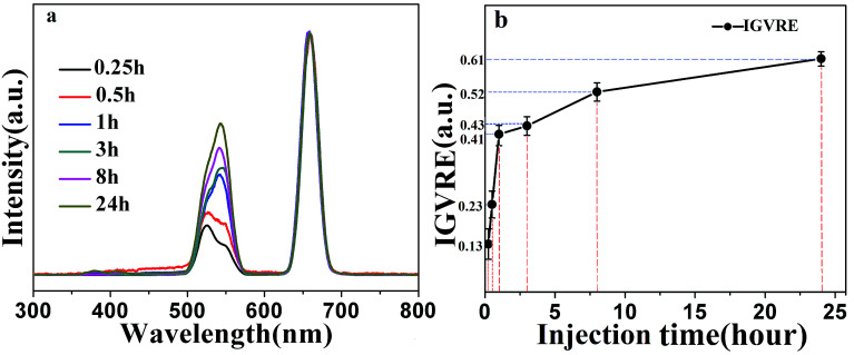
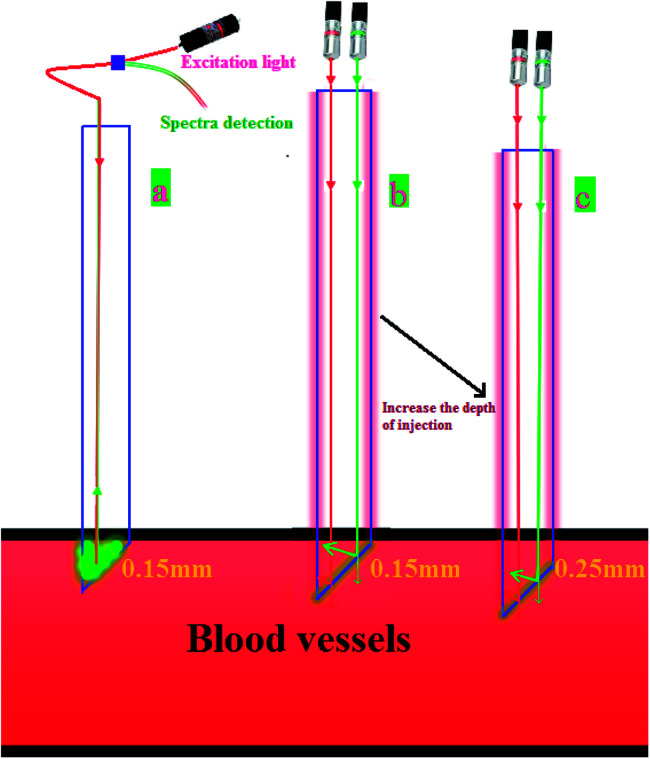
Doxorubicin (Dox) is a chemotherapy medication used to treat cancer. Herein, we report a rapid and efficient method for detecting Dox in vivo based on a NaGdF4:Yb3+,Er3+@NaYF4 core/shell upconversion nanoparticles (UCNPs) probe. We found that the intensity ratio of green to red emission (IGVRE) bands of the core/shell NaGdF4:Yb3+,Er3+@NaYF4 nanoparticles was sensitive to Dox in blood samples, and drops as the concentration of Dox increases. In addition, the proposed UCNPs probe possessed the advantage that no nanoparticles leaked into the living body, thus overcoming the intrinsic defect (difficulty in removing UCNPs from blood vessels) of the fluorescence resonance energy transfer (FRET) approach. This proposed UCNP probe design and results may provide some guidance for the real-time and efficient detection of Dox, and can be helpful in biomedical applications.
This journal is © The Royal Society of Chemistry.
Conflict of interest statement
No conflict of interest exits in the submission of this manuscript, and manuscript is approved by all authors for publication.
Figures
Similar articles
-
Intense Red-Emitting Upconversion Nanophosphors (800 nm-Driven) with a Core/Double-Shell Structure for Dual-Modal Upconversion Luminescence and Magnetic Resonance in Vivo Imaging Applications.ACS Appl Mater Interfaces. 2018 Apr 18;10(15):12331-12340. doi: 10.1021/acsami.7b18078. Epub 2018 Apr 4. ACS Appl Mater Interfaces. 2018. PMID: 29546978
-
Dye-sensitized core-shell NaGdF4:Yb,Er@NaGdF4:Yb,Nd upconversion nanoprobe for determination of H2S.Spectrochim Acta A Mol Biomol Spectrosc. 2021 Mar 5;248:119281. doi: 10.1016/j.saa.2020.119281. Epub 2020 Dec 3. Spectrochim Acta A Mol Biomol Spectrosc. 2021. PMID: 33310610
-
Self-Assembled Upconversion Nanoparticle Clusters for NIR-controlled Drug Release and Synergistic Therapy after Conjugation with Gold Nanoparticles.Inorg Chem. 2017 May 1;56(9):5295-5304. doi: 10.1021/acs.inorgchem.7b00380. Epub 2017 Apr 12. Inorg Chem. 2017. PMID: 28402112
-
Monodisperse Core-Shell NaYF4:Yb3+/Er3+@NaYF4:Nd3+-PEG-GGGRGDSGGGY-NH2 Nanoparticles Excitable at 808 and 980 nm: Design, Surface Engineering, and Application in Life Sciences.Front Chem. 2020 Jun 12;8:497. doi: 10.3389/fchem.2020.00497. eCollection 2020. Front Chem. 2020. PMID: 32596210 Free PMC article.
-
Smart design of exquisite multidimensional multilayered sand-clock-like upconversion nanostructures with ultrabright luminescence as efficient luminescence probes for bioimaging application.Mikrochim Acta. 2020 Aug 28;187(9):527. doi: 10.1007/s00604-020-04521-2. Mikrochim Acta. 2020. PMID: 32860120
Cited by
-
Effect of a Low-Level Laser on Liposomal Doxorubicin Efficacy in a Melanoma Cell Line.J Lasers Med Sci. 2021 Jun 20;12:e28. doi: 10.34172/jlms.2021.28. eCollection 2021. J Lasers Med Sci. 2021. PMID: 34733751 Free PMC article.
-
Highly Red Light-Emitting Erbium- and Lutetium-Doped Core-Shell Upconverting Nanoparticles Surface-Modified with PEG-Folic Acid/TCPP for Suppressing Cervical Cancer HeLa Cells.Pharmaceutics. 2020 Nov 17;12(11):1102. doi: 10.3390/pharmaceutics12111102. Pharmaceutics. 2020. PMID: 33212942 Free PMC article.
-
Construction of a near-infrared excited RhB@UCNPs fluorescent probe for hypochlorite detection.Mikrochim Acta. 2025 Jul 10;192(8):493. doi: 10.1007/s00604-025-07309-4. Mikrochim Acta. 2025. PMID: 40640485
-
ZnS QDs Stabilized Concurrently with Glutathione and L-cysteine for Highly Sensitive Determining Adriamycin Based on the Fluorescence Enhancement Mechanism.J Fluoresc. 2024 Nov;34(6):2523-2531. doi: 10.1007/s10895-023-03452-4. Epub 2023 Oct 13. J Fluoresc. 2024. PMID: 37831353
-
Nanoheterostructures (NHS) and Their Applications in Nanomedicine: Focusing on In Vivo Studies.Materials (Basel). 2019 Jan 3;12(1):139. doi: 10.3390/ma12010139. Materials (Basel). 2019. PMID: 30609839 Free PMC article. Review.
References
-
- Wang F. Han Y. Lim C. S. Lu Y. H. Xu J. et al. Simultaneous phase and size control of upconversion nanocrystals through lanthanide doping. Nature. 2010;463:1061e5. - PubMed
-
- Chen Z. H. Wu X. F. Hu S. G. Hu P. Yan H. Y. Tang Z. J. Liu Y. X. Upconversion NaLuF4 fluorescent nanoprobes for jellyfish cell imaging and irritation assessment of organic dyes. J. Mater. Chem. C. 2015;3:6067–6076. doi: 10.1039/C5TC00802F. - DOI
LinkOut - more resources
Full Text Sources