

doi: 10.1039/c8ra03687j.
eCollection 2018 Jun 8.
The self-assembly of monosubstituted BODIPY and HFBI-RGD
Affiliations
- PMID: 35539954
- PMCID: PMC9080923
- DOI: 10.1039/c8ra03687j
Item in Clipboard
The self-assembly of monosubstituted BODIPY and HFBI-RGD
RSC Adv.
.
Abstract
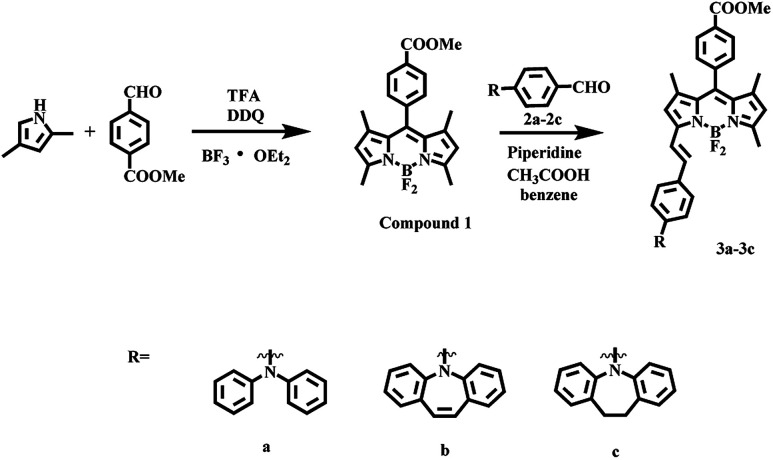
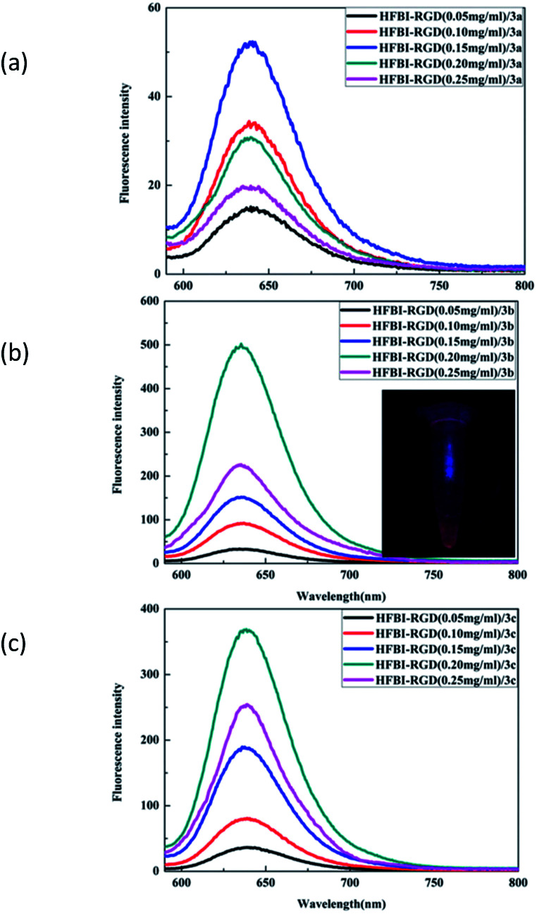
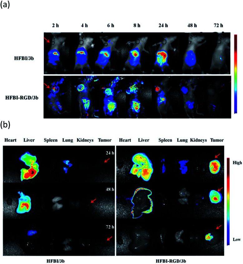
A novel fluorescent probe was constructed by the self-assembly of monosubstituted BODIPY and a novel targeted hydrophobin named hereafter as HFBI-RGD. Optical measurements and theoretical calculations confirmed that the spectral properties of the probe were greatly influenced by the BODIPY structure, the appropriate volume of BODIPY and the cavity of HFBI-RGD. The experiments in vivo and ex vivo demonstrated that the probe had excellent ability for tumor labelling.
This journal is © The Royal Society of Chemistry.
Conflict of interest statement
There are no conflicts to declare.
Figures

Similar articles
-
Dual-functional protein for one-step production of a soluble and targeted fluorescent dye.Theranostics. 2018 Apr 30;8(11):3111-3125. doi: 10.7150/thno.24613. eCollection 2018. Theranostics. 2018. PMID: 29896306 Free PMC article.
-
Lewis acid-assisted isotopic 18F-19F exchange in BODIPY dyes: facile generation of positron emission tomography/fluorescence dual modality agents for tumor imaging.Theranostics. 2013;3(3):181-9. doi: 10.7150/thno.5984. Epub 2013 Feb 21. Theranostics. 2013. PMID: 23471211 Free PMC article.
-
Flattened-Top Domical Water Drops Formed through Self-Organization of Hydrophobin Membranes: A Structural and Mechanistic Study Using Atomic Force Microscopy.ACS Nano. 2016 Jan 26;10(1):81-7. doi: 10.1021/acsnano.5b04049. Epub 2015 Dec 1. ACS Nano. 2016. PMID: 26595357
-
Overproduction, purification, and characterization of the Trichoderma reesei hydrophobin HFBI.Appl Microbiol Biotechnol. 2001 Oct;57(1-2):124-30. doi: 10.1007/s002530100728. Appl Microbiol Biotechnol. 2001. PMID: 11693908
-
Interaction and comparison of a class I hydrophobin from Schizophyllum commune and class II hydrophobins from Trichoderma reesei.Biomacromolecules. 2006 Apr;7(4):1295-301. doi: 10.1021/bm050676s. Biomacromolecules. 2006. PMID: 16602752
Cited by
-
Nonbenzenoid BODIPY Analogues: Synthesis, Structural Organization, Photophysical Studies, and Cell Internalization of Biocompatible N-Alkyl-Aminotroponyl Difluoroboron (Alkyl-ATB) Complexes.ACS Omega. 2022 Aug 1;7(31):27347-27358. doi: 10.1021/acsomega.2c02379. eCollection 2022 Aug 9. ACS Omega. 2022. PMID: 35967069 Free PMC article.
-
Novel near-infrared BODIPY-cyclodextrin complexes for photodynamic therapy.Heliyon. 2024 Feb 23;10(5):e26907. doi: 10.1016/j.heliyon.2024.e26907. eCollection 2024 Mar 15. Heliyon. 2024. PMID: 38449663 Free PMC article.
-
Hydrophobins: multifunctional biosurfactants for interface engineering.J Biol Eng. 2019 Jan 23;13:10. doi: 10.1186/s13036-018-0136-1. eCollection 2019. J Biol Eng. 2019. PMID: 30679947 Free PMC article. Review.
References
-
- Mao M. Li Q.-S. Zhang X.-L. Wu G.-H. Dai C.-G. Ding Y. Dai S.-Y. Song Q.-H. Dyes Pigm. 2017;141:148–160. doi: 10.1016/j.dyepig.2017.02.017. - DOI
-
- Ooyama Y. Kanda M. EnoKi T. Adachi Y. Ohshita J. RSC Adv. 2017;7:13072–13081. doi: 10.1039/C7RA00799J. - DOI
-
- Durán-Sampedro G. Epelde-Elezcano N. Martínez-Martínez V. Esnal I. Bañuelos J. García-Moreno I. Agarrabeitia A. R. de la Moya S. Tabero A. Lazaro-Carrillo A. Villanueva A. Ortiz M. J. López-Arbeloa I. Dyes Pigm. 2017;142:77–87. doi: 10.1016/j.dyepig.2017.03.026. - DOI
LinkOut - more resources
Full Text Sources