. 2019 Nov 26;9(66):38558-38567.
doi: 10.1039/c9ra07622k. eCollection 2019 Nov 25.

Panax notoginseng saponins radiosensitize colorectal cancer cells by regulating the SNHG6/miR-137 axis

Affiliations

Panax notoginseng saponins radiosensitize colorectal cancer cells by regulating the SNHG6/miR-137 axis

Caihui Xu et al. RSC Adv. .

Abstract

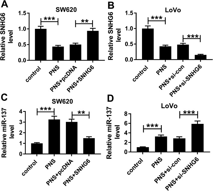
Panax notoginseng saponins (PNS) have recently attracted great attention for their anti-cancer activity in colorectal cancer (CRC). The aim of this study was to explore the functional role and underlying mechanisms of PNS on CRC radiosensitivity. Cell viability was assessed by a Cell Counting kit-8 assay. Cell survival and apoptosis were determined using colony formation assay and flow cytometry, respectively. Quantitative real-time PCR was used to quantify the levels of SNHG6 and miR-137. The targeted correlation between SNHG6 and miR-137 was validated by dual-luciferase reporter and RNA immunoprecipitation assays. Our data supported that PNS weakened the viability of CRC cells. Moreover, PNS promoted the radiosensitivity of CRC cells. Mechanistically, PNS enhanced CRC cell radiosensitivity by upregulating SNHG6. SNHG6 directly targeted miR-137 and inhibited miR-137 expression. MiR-137 was involved in the regulatory effect of SNHG6 on CRC cell radiosensitivity. Furthermore, PNS increased miR-137 expression through SNHG6 in CRC cells. Our study suggested that PNS promoted radiosensitivity in CRC cells at least partly through regulating the SNHG6/miR-137 axis, providing a novel understanding of the anti-cancer mechanism of PNS in CRC.


Conflict of interest statement

There is no conflict of interest regarding the publication of this paper.

Figures

Fig. 1
Fig. 1. PNS inhibited CRC cell viability. Human noncancerous NCM460 colon cells (A), SW620 (B) and LoVo (C) cells were exposed to different concentration (0, 50, 100 and 200 μM) of PNS for different time point (12, 24 and 48 h), followed by the determination of cell viability by CCK-8 assay. (D) The IC50 value for PNS in SW620 and LoVo cells by CCK-8 assay. *P < 0.05, **P < 0.01 or ***P < 0.001.
Fig. 2
Fig. 2. PNS increased CRC cell sensitivity to radiotherapy. SW620 (A) and LoVo (B) cells were exposed to 200 μM of PNS or 0.1% DMSO for 24 h, and then irradiated with increasing dosage intervals (0, 2, 4, 6 and 8 Gy) of X-rays for 24 h, followed by the detection of cell survival using a standard colony formation assay. SW620 (C) and LoVo (D) cells were treated with 200 μM of PNS or 0.1% DMSO for 24 h before radiation with or without 4 Gy of X-rays for 24 h, followed by the determination of cell apoptosis using flow cytometry. *P < 0.05, **P < 0.01 or ***P < 0.001.
Fig. 3
Fig. 3. PNS repressed SNHG6 expression in CRC cells. qRT-PCR for SNHG6 expression in 46 pairs of CRC tissues and adjacent noncancerous tissues (A), in CRC cell lines SW620 and LoVo and noncancerous NCM460 colon cells (B). SW620 (C) and LoVo (D) cells were exposed to 200 μM of PNS for different time point (0, 12, 24 and 48 h), followed by the determination of SNHG6 level by qRT-PCR. *P < 0.05, **P < 0.01 or ***P < 0.001.
Fig. 4
Fig. 4. PNS enhanced radiosensitivity by SNHG6 in CRC cells. (A and B) SW620 and LoVo cells were transfected with or without SNHG6 overexpression plasmid (pcDNA-SNHG6) or negative plasmid pcDNA-con for 24 h prior to 200 μM PNS treatment for 24 h, and then SNHG6 expression was assessed by qRT-PCR. (C and D) SW620 and LoVo cells were transfected with or without pcDNA-SNHG6 or pcDNA-con prior to PNS treatment for 24 h and irradiation with increasing dosage intervals (0, 2, 4, 6 and 8 Gy) of X-rays for 24 h, followed by the measurement of cell survival using a standard colony formation assay. (E and F) SW620 and LoVo cells were transfected with or without pcDNA-SNHG6 or pcDNA-NC before PNS treatment and irradiation with 4 Gy of X-rays, followed by the detection of cell apoptosis by flow cytometry. **P < 0.01 or ***P < 0.001.
Fig. 5
Fig. 5. SNHG6 directly targeted miR-137 and inhibited miR-137 expression. (A) Schematic of the putative miR-137 binding site in SNHG6 and site-directed mutant of seeded sequence. (B and C) Relative luciferase activity in SW620 and LoVo cells cotransfected with SNHG6 luciferase reporter (SNHG6-WT) or its mutant in seeded sequence (SNHG6-MUT) and miR-137 mimic or miR-con mimic. (D and E) The levels of miR-137 and SNHG6 by qRT-PCR in the RISC of SW620 and LoVo cells using anti-Ago2 or IgG antibody. (F) The expression of miR-137 by qRT-PCR in CRC cell lines SW620 and LoVo and noncancerous NCM460 colon cells. (G and H) MiR-137 level in SW620 and LoVo cells transfected with pcDNA-con, pcDNA-SNHG6, si-con or si-SNHG6. ***P < 0.001.
Fig. 6
Fig. 6. MiR-137 was involved in the regulatory effect of SNHG6 on radiosensitivity in CRC cells. SW620 and LoVo cells were transfected with si-con, si-SNHG6, si-SNHG6 + anti-miR-con or si-SNHG6 + anti-miR-137. (A and B) 48 h after transfection, miR-137 level was detected by qRT-PCR. (C and D) After 24 h post-transfection, cells were irradiated with increasing dosage intervals (0, 2, 4, 6 and 8 Gy) of X-rays for 24 h, and then cell survival was assessed using a standard colony formation assay. (E and F) Cells were irradiated with 4 Gy of X-rays for 24 h after transfection for 24 h, and then apoptotic rate was determined by flow cytometry. **P < 0.01 or ***P < 0.001.
Fig. 7
Fig. 7. PNS increased miR-137 expression through SNHG6 in CRC cells. SW620 and LoVo cells were transfected with or without pcDNA-con or pcDNA-SNHG6 for 24 h before treatment with 200 μM of PNS or 0.1% DMSO for 24 h, followed by the determination of SNHG6 level (A and B) and miR-137 expression (C and D) by qRT-PCR. **P < 0.01 or ***P < 0.001.
Fig. 8
Fig. 8. Schematic model of the PNS/SNHG6/miR-137 axis on CRC cell radiosensitivity. PNS reduced SNHG6 expression in CRC cells. Then, miR-137 expression was significantly elevated in SNHG6-silencing CRC cells. Finally, a high miR-137 level enhanced CRC cell radiosensitivity through the regulation of cell viability and apoptosis.

Similar articles

Cited by

References

    1. Bray F. Ferlay J. Soerjomataram I. Siegel R. L. Torre L. A. Jemal A. Ca-Cancer J. Clin. 2018;68:394–424. doi: 10.3322/caac.21492. - DOI - PubMed
    1. Barker H. E. Paget J. T. Khan A. A. Harrington K. J. Nat. Rev. Cancer. 2015;15:409–425. doi: 10.1038/nrc3958. - DOI - PMC - PubMed
    1. Huang E. Y. Chang J. C. Chen H. H. Hsu C. Y. Hsu H. C. Wu K. L. BMC Cancer. 2018;18:321. doi: 10.1186/s12885-018-4254-4. - DOI - PMC - PubMed
    1. Wang X. C. Yue X. Zhang R. X. Liu T. Y. Pan Z. Z. Yang M. J. Lu Z. H. Wang Z. Y. Peng J. H. Le L. Y. Wang G. Y. Peng Q. H. Meng Y. Huang W. Liu R. Y. Clin. Cancer Res. 2019;25:4567–4579. doi: 10.1158/1078-0432.CCR-18-3735. - DOI - PubMed
    1. Yang W. Z. Hu Y. Wu W. Y. Ye M. Guo D. A. Phytochemistry. 2014;106:7–24. doi: 10.1016/j.phytochem.2014.07.012. - DOI - PubMed