

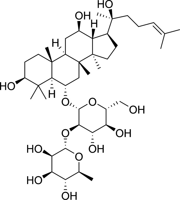
20(S)-Ginsenoside Rg2 attenuates myocardial ischemia/reperfusion injury by reducing oxidative stress and inflammation: role of SIRT1
- PMID: 35540288
- PMCID: PMC9081734
- DOI: 10.1039/c8ra02316f
20(S)-Ginsenoside Rg2 attenuates myocardial ischemia/reperfusion injury by reducing oxidative stress and inflammation: role of SIRT1
Abstract
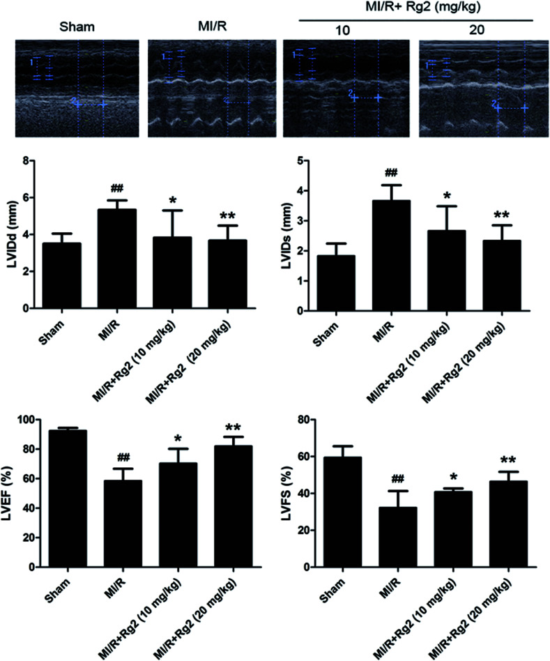
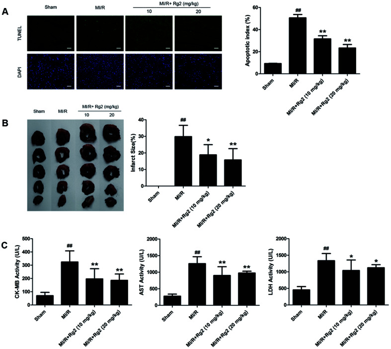
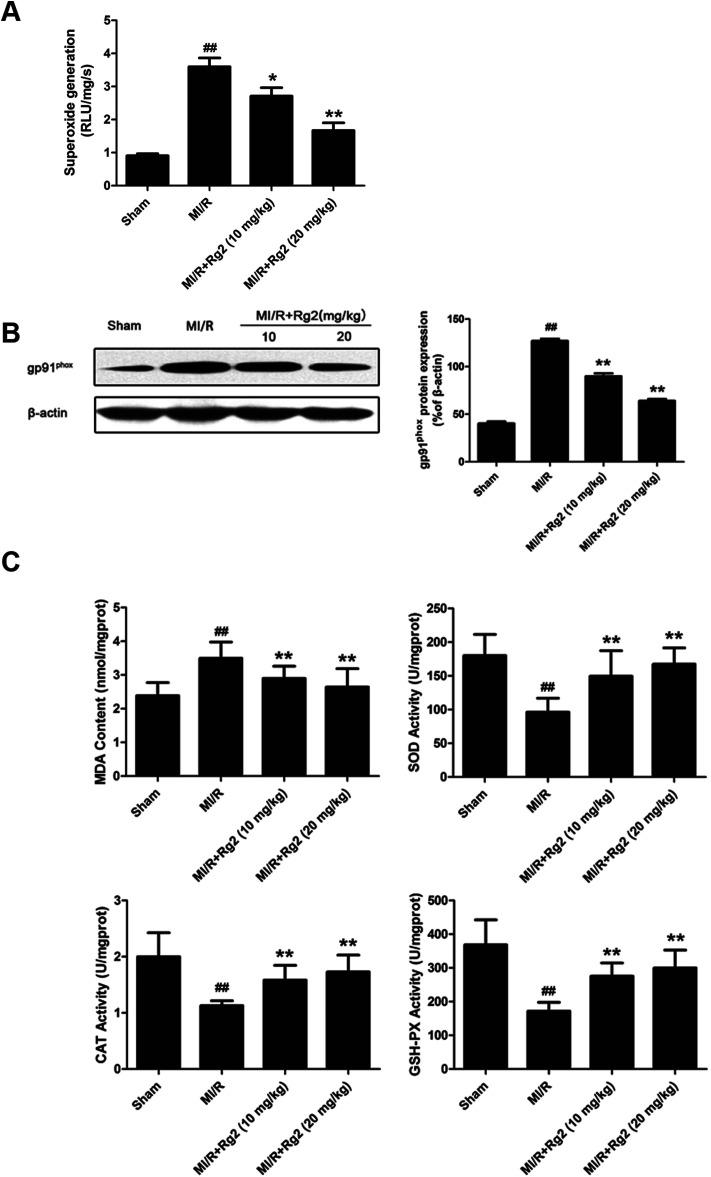
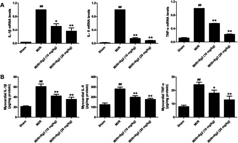
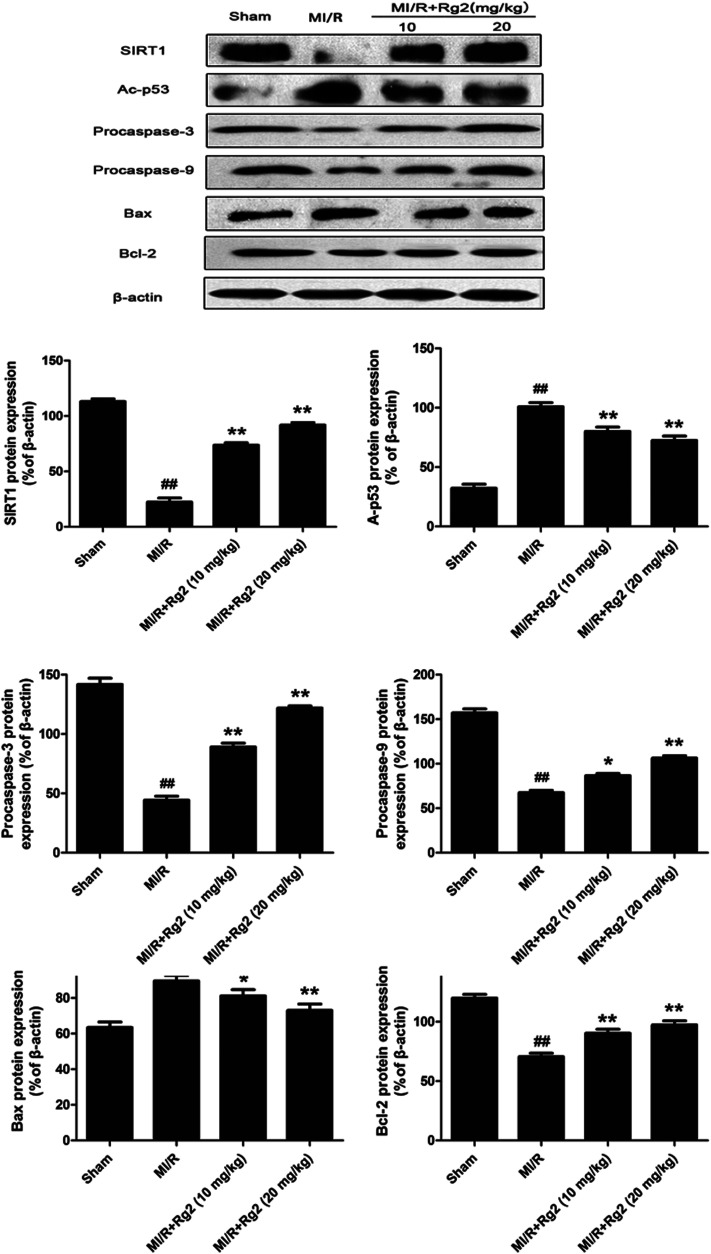
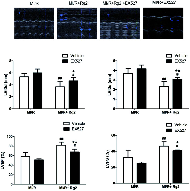
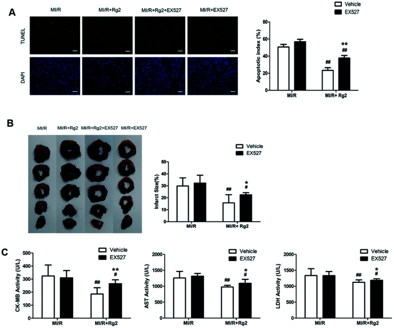
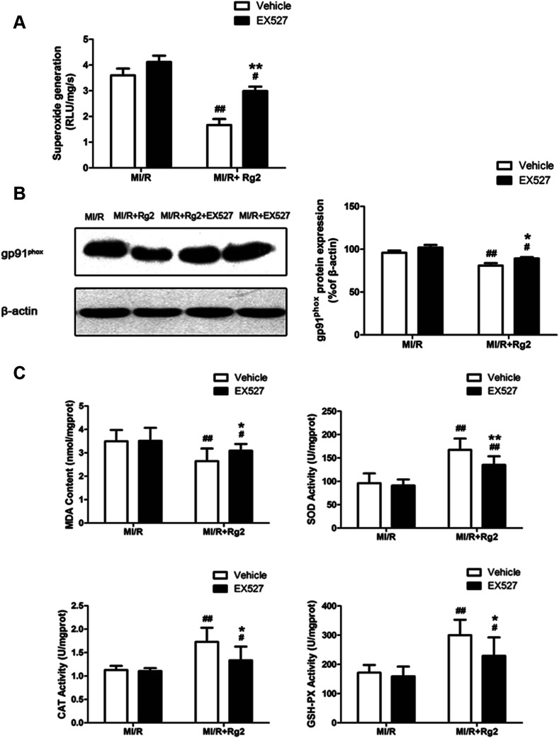
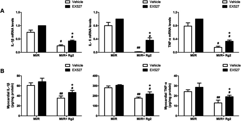
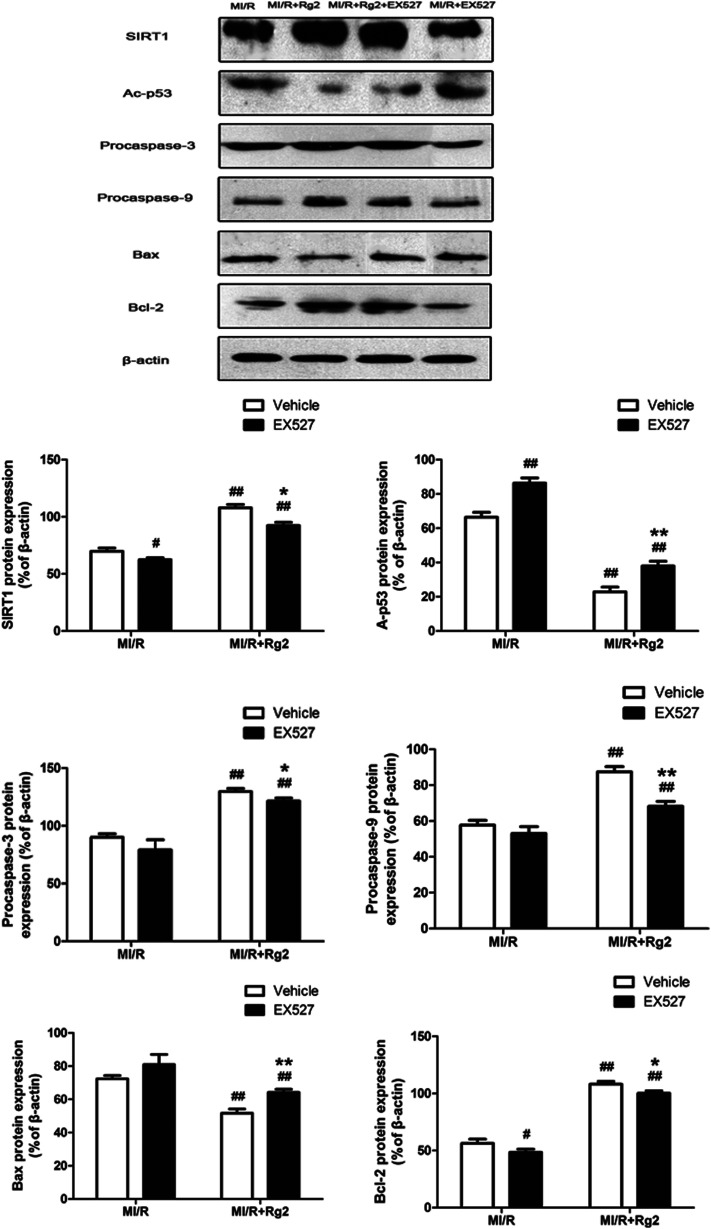
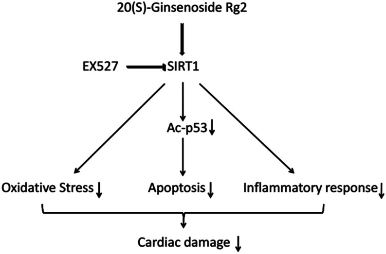
Previously we demonstrated that 20(S)-ginsenoside Rg2 protects cardiomyocytes from H2O2-induced injury by inhibiting reactive oxygen species (ROS) production, increasing intracellular levels of antioxidants and attenuating apoptosis. We explored the protective effect of 20(S)-ginsenoside Rg2 on myocardial ischemia/reperfusion (MI/R) injury and to clarify its potential mechanism of action. Rats were exposed to 20(S)-ginsenoside Rg2 in the presence/absence of the silent information regulator SIRT(1) inhibitor EX527 and then subjected to MI/R. 20(S)-Ginsenoside Rg2 conferred a cardioprotective effect by improving post-ischemic cardiac function, decreasing infarct size, reducing the apoptotic index, diminishing expression of creatine kinase-MB, aspartate aminotransferase and lactate dehydrogenase in serum, upregulating expression of SIRT1, B-cell lymphoma-2, procaspase-3 and procaspase-9, and downregulating expression of Bax and acetyl (Ac)-p53. Pretreatment with 20(S)-ginsenoside Rg2 also resulted in reduced myocardial superoxide generation, gp91phox expression, malondialdehyde content, cardiac pro-inflammatory markers and increased myocardial activities of superoxide dismutase, catalase and glutathione peroxidase. These results suggested that MI/R-induced oxidative stress and inflammation were attenuated significantly by 20(S)-ginsenoside Rg2. However, these protective effects were blocked by EX527, indicating that SIRT1 signaling may be involved in the pharmacological action of 20(S)-ginsenoside Rg2. Our results demonstrated that 20(S)-ginsenoside Rg2 attenuates MI/R injury by reducing oxidative stress and inflammatory responses via SIRT1 signaling.
This journal is © The Royal Society of Chemistry.
Conflict of interest statement
The authors declare no conflict of interests.
Figures
References
LinkOut - more resources
Full Text Sources
Research Materials
Miscellaneous