. 2018 Feb 9;8(12):6460-6470.
doi: 10.1039/c7ra13062g. eCollection 2018 Feb 6.

Design of triphasic poly(lactic- co-glycolic acid) nanoparticles containing a perfluorocarbon phase for biomedical applications

Affiliations

Design of triphasic poly(lactic- co-glycolic acid) nanoparticles containing a perfluorocarbon phase for biomedical applications

Edyta Swider et al. RSC Adv. .

Abstract

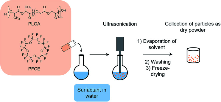
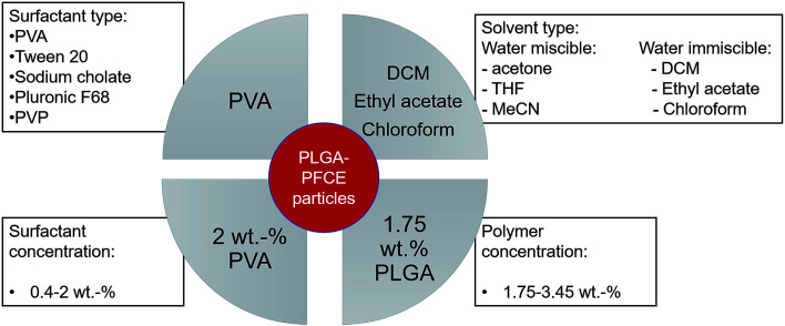
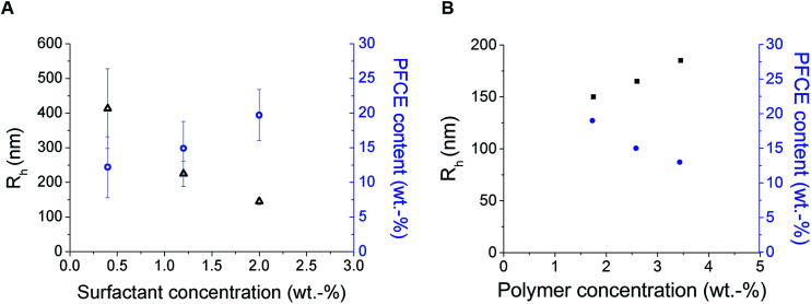
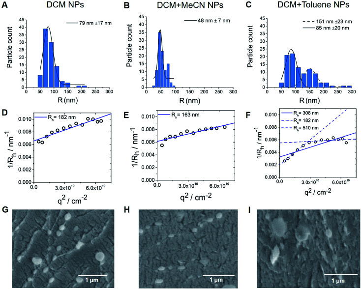
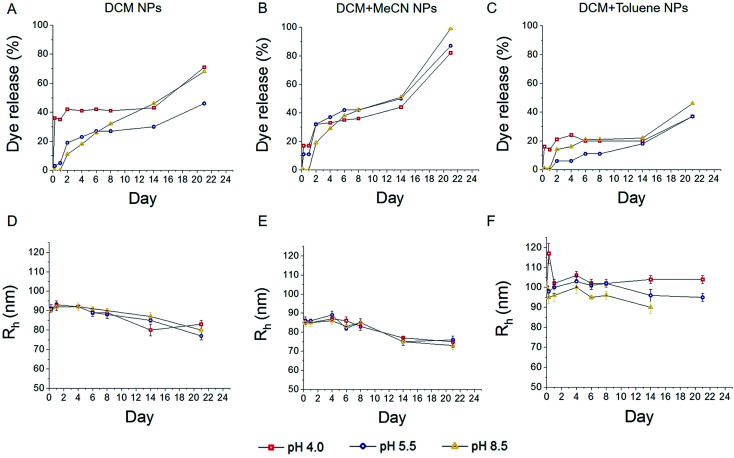
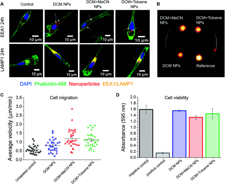
Poly(lactic-co-glycolic acid) (PLGA) particles are very widely used, particularly for drug delivery, including commercial clinical formulations. Adding perfluorocarbon (PFC) enables in vivo imaging and quantification of the PLGA particles through 19F NMR, MRS or MRI. PFCs are both hydrophobic and lipophobic at the same time. This property makes their encapsulation in particles challenging, as it requires the addition of a third immiscible phase during the emulsification process. Here we explore how different parameters affect the miniemulsion formation of particles loaded with perfluoro-15-crown-5-ether (PFCE). By changing the concentration of surfactant and type of solvent, we were able to control the radius of synthesized particles, between 85-200 nm. We assessed stability and release from the particles at different pH values, showing that hydrophobic agents are released from the particles by diffusion rather than degradation. With cell experiments, we show that primary human dendritic cells take up the particles without any apparent effect, including on cell migration. In summary, the control of synthesis conditions leads to particles with sufficient PFCE encapsulation, which are suitable for drug loading and cell labeling, and do not affect cell viability or functionality. Finally, these nanoparticles can be produced at GMP-grade for clinical use.


Conflict of interest statement

There are no conflicts of interest to declare.

Figures

Fig. 1
Fig. 1. Schematic representation of miniemulsion formulation method. PLGA and PFCE are added to surfactant solution under sonication, followed by solvent evaporation. Next, particles are washed, freeze-dried, and collected as a dry powder.
Scheme 1
Scheme 1. Schematic representation of parameters, which affect properties of nanoparticles, tested in this study, including factors that resulted in nanoparticles with low PDI, narrow size distribution and high PFCE content.
Fig. 2
Fig. 2. Results of DLS (black) and NMR (blue) measurements of nanoparticles prepared with different concentration of surfactant (A) or PLGA (B). (A) Influence of PVA concentration on particle radius and PFCE encapsulation. Particles were prepared using following concentrations of surfactant: 0.4 wt%, 1.2 wt% and 2 wt%. Here, the concentration of polymer in organic phase was the same for all samples, 1.75 wt%. Better reproducibility of particle radius was achieved with higher concentration of surfactant. Furthermore, higher concentration of PVA resulted in nanoparticles with higher encapsulation efficiency of PFCE. Error bars represent the standard deviation (n = 3) (B) influence of concentration of PLGA on particle radius and PFCE encapsulation. Higher concentration of polymer resulted in slightly larger particles, but lower encapsulation efficiency. For all samples concentration of surfactant at 2 wt% was used. DLS results are presented in black and NMR results in blue. DLS: c(NP) = 0.1 mg ml−1, NMR with TFA as internal reference.
Fig. 3
Fig. 3. (A–C) Nanoparticle size distribution based on TEM images. Bimodal size distribution can be observed for particles made with the combination of DCM with toluene solvent (3C). (D–F) Multi-angle LS results, where extrapolation q → 0 results in inverse z-average radii <Rh>1/z of 182, 163 and 308 nm for DCM, DCM + MeCN and DCM + toluene nanoparticles respectively. (G–I) CryoSEM images, here both fractions of particles are visible for particles made with DCM + toluene (cNP = 10 mg ml−1). Also, some larger particles could be found with samples made either with DCM only or with DCM + MeCN solvents.
Fig. 4
Fig. 4. Cumulative dye release and changes in particle diameter after incubation at different pH. Faster dye release was observed for smallest particles (B), when compared to particles emulsified with DCM solvent (A), while larger particles resulted in slower release (C). DLS analysis of incubated samples, showed a slow gradual decrease in particles radius (D–F). Lines indicate trend of the data. NPs = nanoparticles.
Fig. 5
Fig. 5. Effect of nanoparticles on monocyte-derived DCs. (A) Confocal microscope images of nanoparticle uptake by DCs. Fluorescent signal coming from the nanoparticles partially overlaps with the signal of the early endosomal marker EEA1. Higher colocalization of nanoparticles was observed with the signal of late endosomal/lysosomal marker LAMP1. (B) 1H/19F MRI image showing the signal of 5 × 10⁶ cells loaded with different nanoparticles. Here, PFCE–PLGA particles, at 10 mg ml−1 concentration, were used as a reference. (C) Nanoparticles do not affect the migration of dendritic cells. The graph represents average velocity of unloaded cells and cells loaded with nanoparticles. (D) The graph shows the viability of cells after incubation with various nanoparticles for 72 hours. No significant influence on cell viability was observed. Error bars represent the standard deviation (n = 3).

Similar articles

Cited by

References

    1. Fuchs A. V. Bapat A. P. Cowin G. J. Thurecht K. J. Switchable 19F MRI polymer theranostics: towards in situ quantifiable drug release. Polym Chem. 2017;8(34):5157–5166. doi: 10.1039/C7PY00345E. - DOI
    1. Kumari A. Yadav S. K. Yadav S. C. Biodegradable polymeric nanoparticles based drug delivery systems. Colloids Surf., B. 2010;75(1):1–18. doi: 10.1016/j.colsurfb.2009.09.001. - DOI - PubMed
    1. Srinivas M. Cruz L. J. Bonetto F. Heerschap A. Figdor C. G. et al., Customizable, multi-functional fluorocarbon nanoparticles for quantitative in vivo imaging using 19F MRI and optical imaging. Biomaterials. 2010;31(27):7070–7077. doi: 10.1016/j.biomaterials.2010.05.069. - DOI - PubMed
    1. Srinivas M. Boehm-Sturm P. Figdor C. G. de Vries I. J. M. Hoehn M. Labeling cells for in vivo tracking using (19)F MRI. Biomaterials. 2012;33(34):8830–8840. doi: 10.1016/j.biomaterials.2012.08.048. - DOI - PubMed
    1. Tel J. Sittig S. P. Blom R. A. M. Cruz L. J. Schreibelt G. et al., Targeting uptake receptors on human plasmacytoid dendritic cells triggers antigen cross-presentation and robust type I IFN secretion. J. Immunol. 2013;191(10):5005–5012. doi: 10.4049/jimmunol.1300787. - DOI - PubMed