. 2022 May 3:30:101265.
doi: 10.1016/j.bbrep.2022.101265. eCollection 2022 Jul.

Piezoelectric effect stimulates the rearrangement of chondrogenic cells and alters ciliary orientation via atypical PKCζ

Affiliations

Piezoelectric effect stimulates the rearrangement of chondrogenic cells and alters ciliary orientation via atypical PKCζ

Jormay Lim et al. Biochem Biophys Rep. .

Abstract

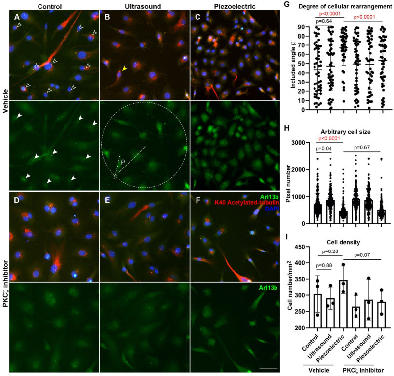
Therapeutic ultrasound was administered to patients suffering from bone fracture with FDA approval. Bone and cartilage are piezoelectric materials. To investigate the effects of piezoelectricity on the cells of chondrogenic lineage, we applied ultrasound stimulation on an AT-cut quartz coverslip to generate electric field fluctuations. The bone-marrow-derived mesenchymal stem cells (BMMSC) and primary chondrocytes were cultured on either glass or quartz coverslips for ultrasound stimulation. The cells were immunofluorescent-labeled for the assessment of cell arrangement and ciliary orientation. Ultrasound and piezoelectricity both stimulate cell migration and disrupt ciliary orientation induced by directional migration. In particular, piezoelectric effects on cell rearrangement can be abolished by the inhibitor specifically targeting atypical Protein kinase C zeta (PKCζ). Our findings shed light on the possibility of cellular modulation by using piezoelectric manipulation.

Keywords: Cell rearrangement; Chondrogenic cells; Ciliary orientation; PKCζ; Piezoelectric stimulation.


Conflict of interest statement

The authors declare that they have no known competing financial interests or personal relationships that could have appeared to influence the work reported in this paper.

Figures

Fig. 1
Fig. 1
Experimental device, method, and estimated magnitude of electrical field for piezoelectric stimulation. (A) Mesh diagram generated for a 3D model of our experimental setup. (B) Electric field distribution on the coverslip. (C) Electric field across a cross section of the quartz coverslip. The results derived from this graph indicated that the electrical field measurement on the cover slips was 1mV/100 μm. (D) Schematic diagram illustrating the electrical field gradient experienced by a cell during piezoelectric stimulation. The dynamic electrical field promotes cell migration and rearranges cell polarity.
Fig. 2
Fig. 2
Piezoelectric stimulation induces cellular rearrangement of BMMSC. Micrograph of BMMSC in stem cell media under control condition (A), ultrasound stimulation (B), and piezoelectric stimulation (C). Chondrogenic media cultured BMMSC under control (D), ultrasound stimulation (E), and piezoelectric stimulation (F). (G) Quantification of primary cilia incidence. (H) Quantification of primary cilia size. (I) Schematic diagram illustrating the analysis of cilia/cell alignment. (J) Quantification of cilia/cell orientation alignment. (K) R2 analysis with respect to perfect alignment. These results indicated that the incidence and size of cilia are not significantly altered upon piezoelectric stimulations whereas ciliary position and cell polarity are affected in a statistically significant manner. On the contrary, ultrasound stimulation does not cause such cellular rearrangement.
Fig. 3
Fig. 3
Disruption of wound-space-induced ciliary re-positioning by ultrasound and piezoelectric stimulations. Micrographs of BMMSC in a wound healing assay initially in control (A, B) and 16 h later in control (C, D), ultrasound (E, F), and piezoelectric stimulation (G, H). (I) Comparison of the wound healing percentage normalized to control. (J) Ciliary position relative to nucleus with respect to the wound direction in different experimental groups. The results in A, B, C, D indicate that ciliary position is induced to direct cell migration towards the cell-free area. The results in E, F, G, H indicate that both ultrasound and piezoelectric override this ciliary orientation towards the cell-free area. Counterintuitively, the randomized ciliary position accelerates the migration.
Fig. 4
Fig. 4
Cellular rearrangement of primary chondrocytes can be abolished by the inhibition of PKCζ. Micrographs of chondrocytes in regular control (A), ultrasound (B) and piezoelectric stimulation (C), and with ZIP, PKCζ inhibitor among control (D), ultrasound (E), and piezoelectric stimulation (F). (G) Quantification of cellular orientation with respect to the radial direction of a defined circle separating the cells that are grouped into clusters, and arbitrary circle in the case of control and ultrasound. (H) Quantification of cell sizes. (I) Quantification of cell densities. The piezoelectric induced cellular rearrangement effect is also observed in chondrocytes. This rearrangement is abolished if pseudosubstrate of PKCζ is added into the media during the piezoelectric stimulation.

Similar articles

Cited by

References

    1. Uddin S.M.Z., Komatsu D.E., Motyka T., Petterson S. Low-intensity continuous ultrasound therapies-A systemic review of current state-of-the-art and future perspectives. J. Clin. Med. 2021;10 doi: 10.3390/jcm10122698. - DOI - PMC - PubMed
    1. Chen C., Zhang T., Liu F., Qu J., Chen Y., Fan S., Chen H., Sun L., Zhao C., Hu J., Lu H. Effect of low-intensity pulsed ultrasound after autologous adipose-derived stromal cell transplantation for bone-tendon healing in a rabbit model. Am. J. Sports Med. 2019;47:942–953. doi: 10.1177/0363546518820324. - DOI - PubMed
    1. Liu D.D., Ullah M., Concepcion W., Dahl J.J., Thakor A.S. The role of ultrasound in enhancing mesenchymal stromal cell-based therapies. Stem Cells Transl. Med. 2020;9:850–866. doi: 10.1002/sctm.19-0391. - DOI - PMC - PubMed
    1. Bloebaum R.D., Wilson A.S., Martin W.N. A review of the collagen orientation in the articular cartilage. Cartilage. 2021 doi: 10.1177/1947603520988770. 1947603520988770. - DOI - PMC - PubMed
    1. Hughes L.C., Archer C.W., ap Gwynn I. The ultrastructure of mouse articular cartilage: collagen orientation and implications for tissue functionality. A polarised light and scanning electron microscope study and review. Eur. Cell. Mater. 2005;9:68–84. doi: 10.22203/ecm.v009a09. - DOI - PubMed

LinkOut - more resources