. 2018 Mar 15;8(19):10509-10515.
doi: 10.1039/c8ra01260a. eCollection 2018 Mar 13.

Layer-by-layer decoration of MOFs on electrospun nanofibers

Affiliations

Layer-by-layer decoration of MOFs on electrospun nanofibers

Jinhong Shangguan et al. RSC Adv. .

Abstract

The design and fabrication of novel organic-inorganic nanocomposite membranes using metal-organic frameworks as building blocks have attracted numerous scientists. Here, HKUST-1 particles were decorated on crosslinked polymer nanofibers through a layer-by-layer method. The immersion sequence, the crosslinking and the number of the deposition cycles have a significant impact on the formation of the HKUST-1 decorated nanofibrous membranes. Moreover, it has been shown that such a membrane could be applied as a catalyst for visual detection of hydrogen peroxide.


Conflict of interest statement

There are no conflicts to declare.

Figures
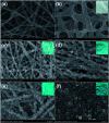
Fig. 1
Fig. 1. SEM images of the pristine PAA/PVA nanofibers (a), the crosslinked PAA/PVA nanofibers (b), MOF-decorated nanofibers after 1 cycle of growth (c), MOF-decorated nanofibers after 2 cycle of growth (d), MOF-decorated nanofibers after 4 cycle of growth (e) and MOF-decorated nanofibers after 8 cycle of growth (f). The insets are the corresponding photos of the membranes.
Fig. 2
Fig. 2. XRD patterns (a), FTIR spectra (b) and TG curves (c) of the PAA/PVA nanofibers, the HKUST-1 powder and the HKUST-1 decorated nanofibers after different cycles of growth.
Fig. 3
Fig. 3. SEM images of various nanofiber membranes. (a) The nanofibers were immersed only into the metal ion solution for 2 h; (b) the nanofibers were immersed into the ligand solution first and then into the metal ion solution; the decoration of HKUST-1 on the nanofibers after crosslinking for 30 min (c) and 1 h (d), and the growth process was identical with the sample in Fig. 1c (after crosslinking for 15 min).
Fig. 4
Fig. 4. UV-vis spectra of the solutions consisted of (a) OPD and H2O2, (b) HKUST-1 decorated nanofiber membrane and H2O2, (c) OPD, (d) OPD and HKUST-1 decorated nanofiber membrane, (e) OPD, H2O2 and HKUST-1 decorated nanofiber membrane. The inset is the corresponding photo of the solutions.
Fig. 5
Fig. 5. (a) UV-vis spectrum of the mixed solution of OPD and H2O2 after the addition of 100 μL of 1 mM Cu(OAc)2 solution; (b) the peak intensity at 418 nm was recorded to test the recyclability of the HKUST-1 decorated nanofiber membrane for the detection of H2O2.

Similar articles

Cited by

References

    1. Stock N. Biswas S. Chem. Rev. 2012;112:933–969. doi: 10.1021/cr200304e. - DOI - PubMed
    1. Furukawa S. Reboul J. Diring S. Sumida K. Kitagawa S. Chem. Soc. Rev. 2014;43:5700–5734. doi: 10.1039/C4CS00106K. - DOI - PubMed
    1. Qiu S. L. Xue M. Zhu G. S. Chem. Soc. Rev. 2014;43:6116–6140. doi: 10.1039/C4CS00159A. - DOI - PubMed
    1. Furukawa H. Cordova K. E. O'Keeffe M. Yaghi O. M. Science. 2013;341:974. doi: 10.1126/science.1230444. - DOI - PubMed
    1. Greiner A. Wendorff J. H. Angew. Chem., Int. Ed. 2007;46:5670–5703. doi: 10.1002/anie.200604646. - DOI - PubMed