. 2018 May 9;8(30):16973-16990.
doi: 10.1039/c8ra00954f. eCollection 2018 May 3.

Copper complexes as prospective anticancer agents: in vitro and in vivo evaluation, selective targeting of cancer cells by DNA damage and S phase arrest

Affiliations

Copper complexes as prospective anticancer agents: in vitro and in vivo evaluation, selective targeting of cancer cells by DNA damage and S phase arrest

Dharmasivam Mahendiran et al. RSC Adv. .

Abstract

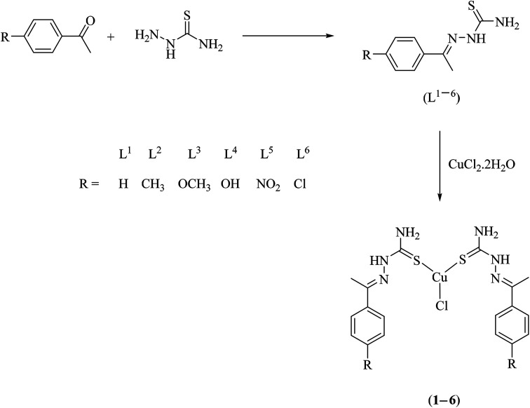
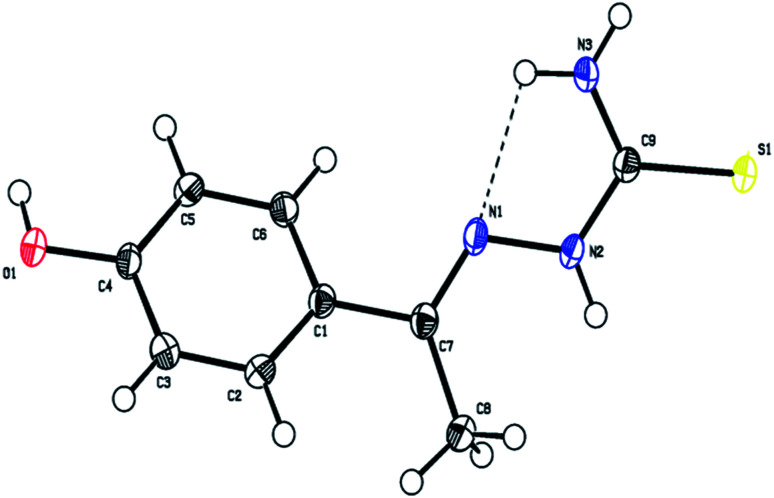
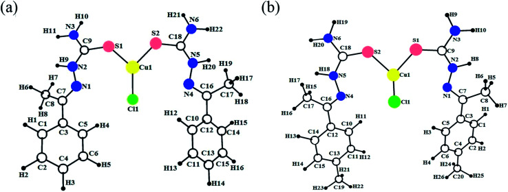
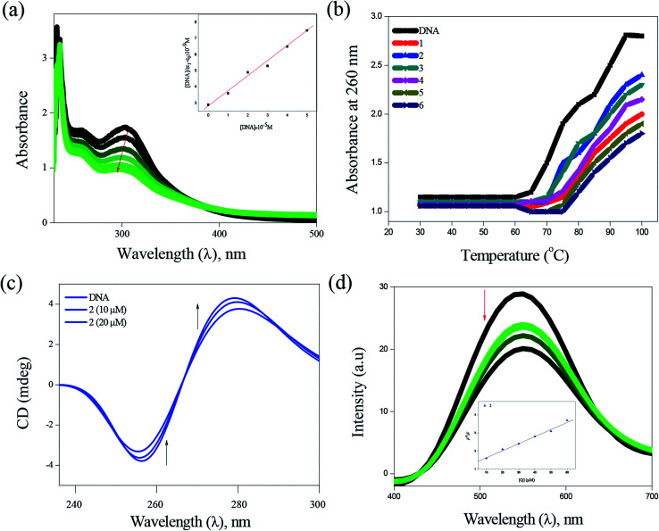
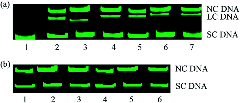
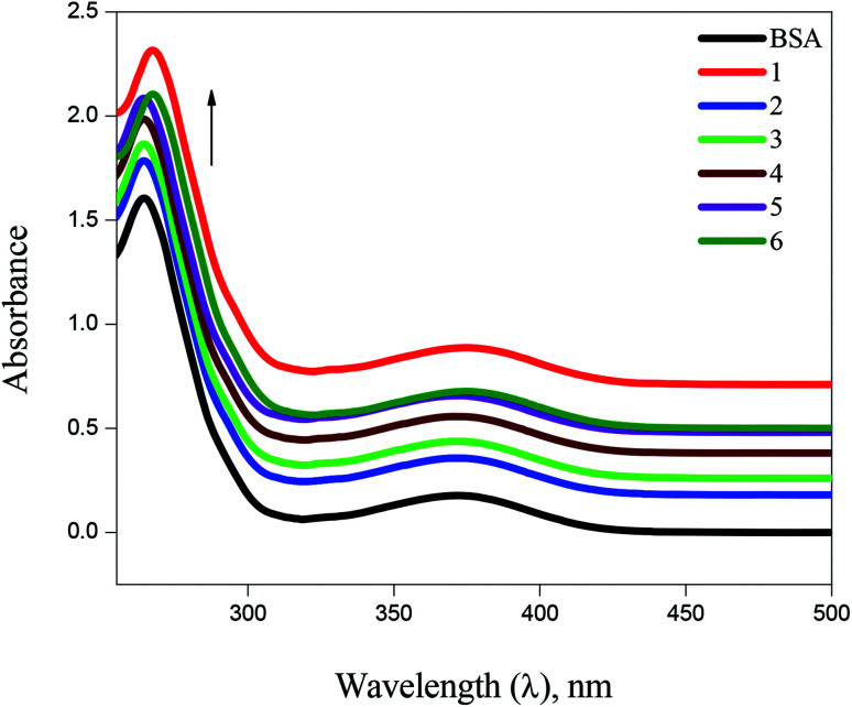
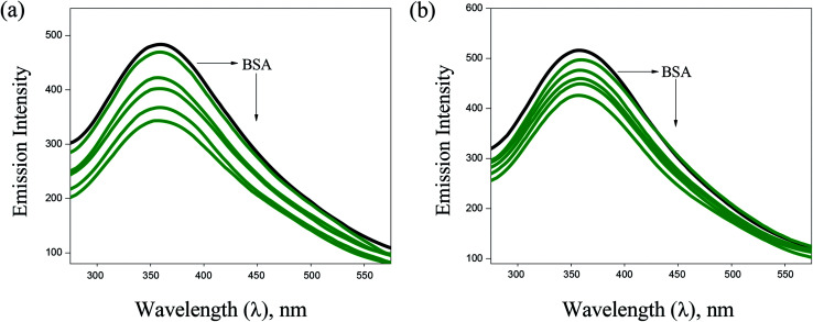
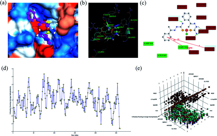
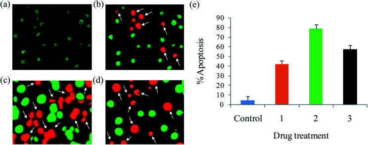
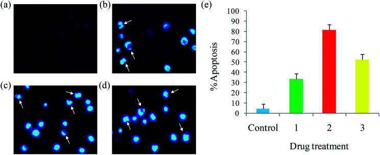
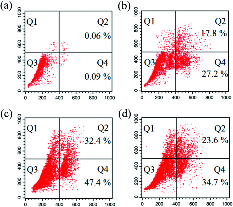
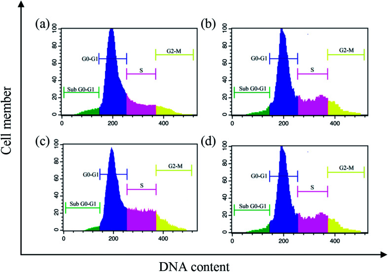
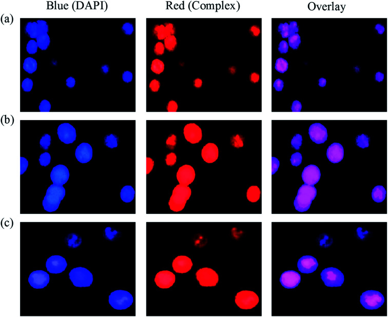
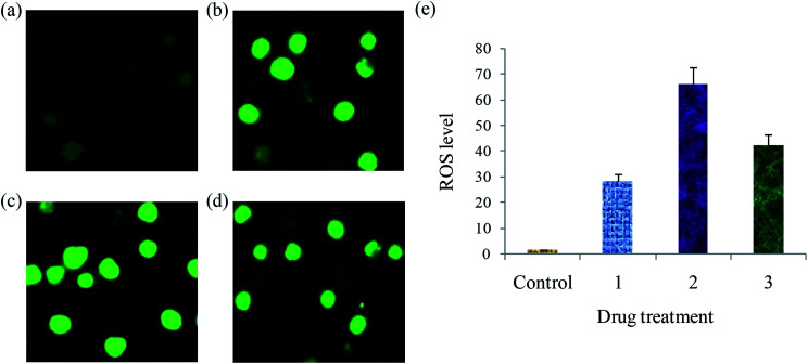
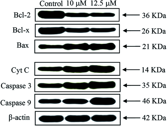
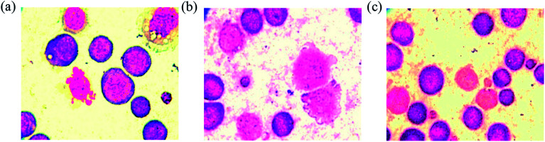
A series of six new bis(thiosemicarbazone)copper(i) complexes of the type [Cu(L1-6)2Cl] (1-6) have been synthesized and characterized. The molecular structure of the ligand L4 was determined by the single crystal XRD method. All the complexes adopted trigonal planar (Y-shaped) geometry. All the complexes strongly bind with CT-DNA via intercalative mode, which was further supported by molecular docking studies. Further, the complexes were effectively bind with BSA as observed by UV-Vis and fluorescence spectra. All the complexes effectively cleave pBR322 DNA through hydrolytic pathway as evidenced from T4 ligase experiments. All the complexes interact with the anticancer receptor focal adhesion kinase (FAK) via electrostatic, van der Waals, hydrogen bonding, σ-π and π-π interactions. In vitro cytotoxicity of the complexes were assessed by MTT assay against four cancer cell lines such as human breast adenocarcinoma (MCF-7), cervical (HeLa), epithelioma (Hep-2) and Ehrlich ascites carcinoma (EAC), and two normal cell lines namely normal human dermal fibroblasts (NHDF) and L6 myotubes with respect to the commercially used anticancer drug cisplatin. All the complexes induce apoptosis in EAC cells, which was confirmed by AO/EB, Hoechst 33258 and PI staining methods. The complexes block cell cycle progression of EAC cells in S phase (DNA synthesis). The cellular uptake studies confirmed the ability of the complexes to go into the cytoplasm and accumulation in the cell nuclei. In the in vivo anticancer studies, the complexes significantly reduce the tumour volume in female Swiss albino mice. Overall, our results ensure the role of thiosemicarbazone-based copper(i) complexes as prospective anticancer agents, induction of apoptosis and S phase arrest with the mitochondrial controlled pathway.


Conflict of interest statement

There are no conflicts to declare.

Figures

Scheme 1
Scheme 1. Synthesis of thiosemicarbazone ligands (L1–6) and their copper(i) complexes (1–6).
Fig. 1
Fig. 1. ESI mass spectrum of complex 2.
Fig. 2
Fig. 2. The molecular structure of the ligand L4 showing the atom labelling scheme. The displacement ellipsoids are drawn at 50% probability level.
Fig. 3
Fig. 3. Optimized geometries of the copper(i) complexes 1 (a) and 2 (b).
Fig. 4
Fig. 4. DNA binding of complexes with CT-DNA: absorption spectra of complex 2 (a), thermal denaturation of complexes 1–6 (b), CD spectra of 2 (c) and emission spectra of complex 2 (d).
Fig. 5
Fig. 5. Hydrolytic cleavage of complexes (1–6) on pBR322 DNA (33.3 μM) in Tris–HCl/NaCl buffer. Lane 1, DNA alone, lane 2–7, DNA + 1–6 (25 μM) (a). Analysis of the capacity of T4 DNA ligase to relegate DNA cleaved by complexes 1–6. Lane 1–6, NC obtained using complexes 1–6 + T4 ligase (b).
Fig. 6
Fig. 6. Absorbance titrations of the complexes (1–6) with BSA.
Fig. 7
Fig. 7. Fluorescence spectra of BSA (10 μM, λex = 280 nm, λem = 570 nm) in the presence of increasing concentrations of 2 (a) and 3 (b). Arrow shows that the emission intensity changes upon increasing the concentration of the complexes.
Fig. 8
Fig. 8. Molecular docking view of complex 1: complex located in hydrophobic packet (subdomains II) (a), 3D interaction (b), 2D interaction (c), hydrophobic interaction plot (d), and 3D interaction plot of residue count (e), with focal adhesion kinase (FAK) receptor.
Fig. 9
Fig. 9. AO/EB staining of EAC cells for 24 h: control (a), complexes 1 (b), 2 (c) and 3 (d), and scar bar diagram (e).
Fig. 10
Fig. 10. Hoechst 33258 staining of EAC cells for 24 h: control (a), complexes 1 (b), 2 (c) and 3 (d), and scar bar diagram (e).
Fig. 11
Fig. 11. Apoptosis detection in EAC cells using the PI assay after 24 h: control (a), and complexes 1 (b) 2 (c) and 3 (d). Four areas in the diagrams represent four different cell states: necrotic cells (Q1), late apoptotic cells (Q2), living cells (Q3) and early apoptotic cells (Q4).
Fig. 12
Fig. 12. Cell cycle progression of EAC cells treated with complexes after 24 h: control (a), and complexes 1 (b) 2 (c) and 3 (d).
Fig. 13
Fig. 13. Images of EAC cell emergence to complexes 1 (a) 2 (b) and 3 (c), stained with DAPI at 37 °C for 24 h.
Fig. 14
Fig. 14. ROS generation of EAC cells after treatment with complexes for 24 h: control (a), complexes 1 (a) 2 (c) and 3 (d), and scar bar diagram (e).
Fig. 15
Fig. 15. Western blot analysis of Bcl-2 family proteins, caspases 3/9 and Cyt C in EAC cells treated with complex 2 for 24 h. β-Actin was used as internal loading control.
Fig. 16
Fig. 16. Smear showing matured EAC control cells with definite structure and clear cell wall without degeneration (a), EAC tumor cells treated with the complexes 2 (b) and 3 (c) showing degenerative changes like membrane blebbing, cell wall destruction, cell disintegration and reduction in staining intensity.

References

    1. Whitnall M. Howard J. Ponka P. Richardson D. R. A class of iron chelators with a wide spectrum of potent antitumor activity that overcomes resistance to chemotherapeutics. Proc. Natl. Acad. Sci. U. S. A. 2006;103:14901–14906. doi: 10.1073/pnas.0604979103. - DOI - PMC - PubMed
    1. Stacy A. E. Palanimuthu D. Bernhardt P. V. Kalinowski D. S. Jansson P. J. Richardson D. R. Zinc(ii)–thiosemicarbazone complexes are localized to the lysosomal compartment where they transmetallate with copper ions to induce cytotoxicity. J. Med. Chem. 2016;59:4965–4984. doi: 10.1021/acs.jmedchem.6b00238. - DOI - PubMed
    1. http://www.clinicaltrials.gov. http://www.clinicaltrials.gov
    1. Vavere A. L. Lewis J. S. Cu-ATSM: a radiopharmaceutical for the PET imaging of hypoxia. Dalton Trans. 2007:4893–4902. doi: 10.1039/B705989B. - DOI - PubMed
    1. Crouch P. J. Hung L. W. Adlard P. A. Cortes M. Lal V. Filiz G. Perez K. A. Nurjono M. Caragounis A. Du T. Laughton K. Volitakis I. Bush A. I. Li Q. X. Masters C. L. Cappai R. Cherny R. A. Donnelly P. S. White A. R. Barnham K. J. Increasing Cu bioavailability inhibits Aβ oligomers and tau phosphorylation. Proc. Natl. Acad. Sci. U. S. A. 2009;106:381–386. doi: 10.1073/pnas.0809057106. - DOI - PMC - PubMed