. 2018 Mar 6;8(18):9603-9610.
doi: 10.1039/c8ra00849c. eCollection 2018 Mar 5.

Creating tissue on chip constructs in microtitre plates for drug discovery

Affiliations

Creating tissue on chip constructs in microtitre plates for drug discovery

N P Macdonald et al. RSC Adv. .

Abstract

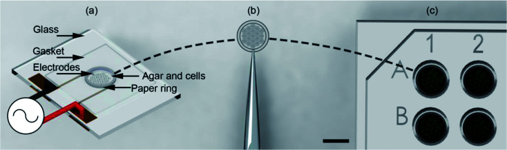
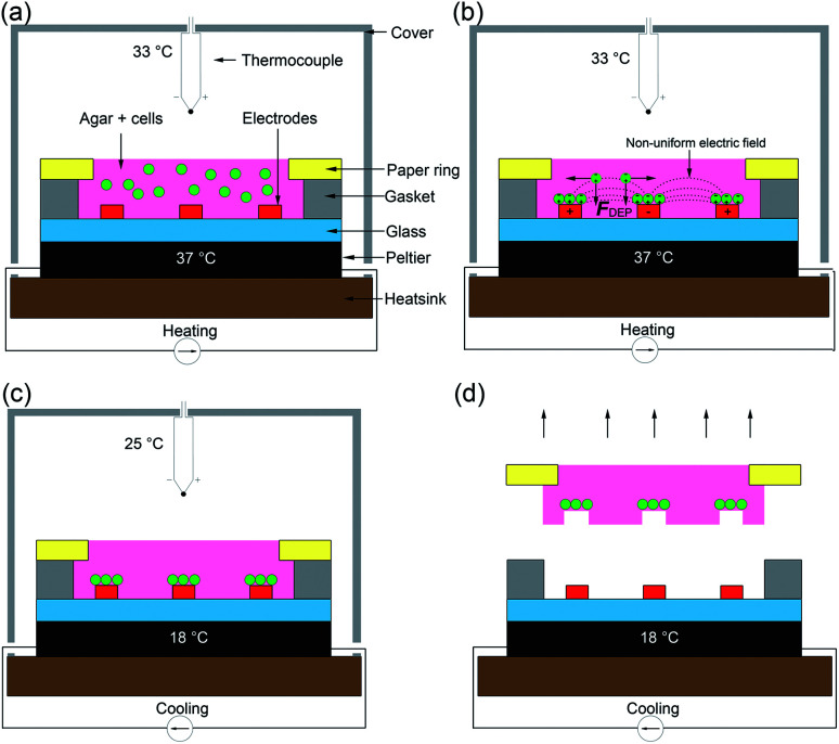
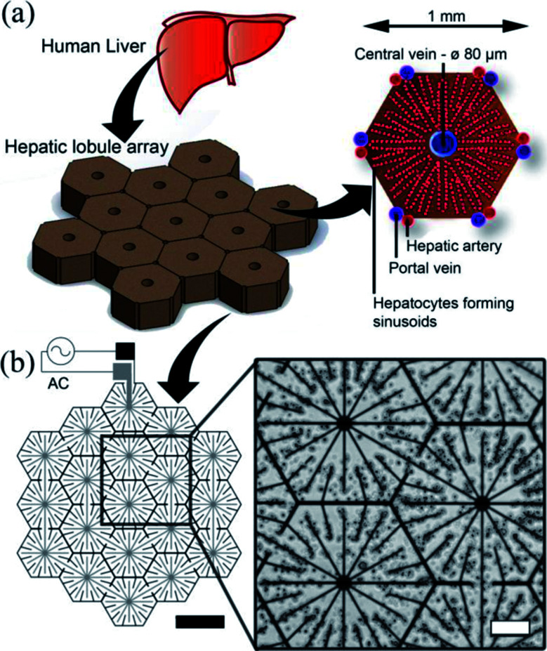
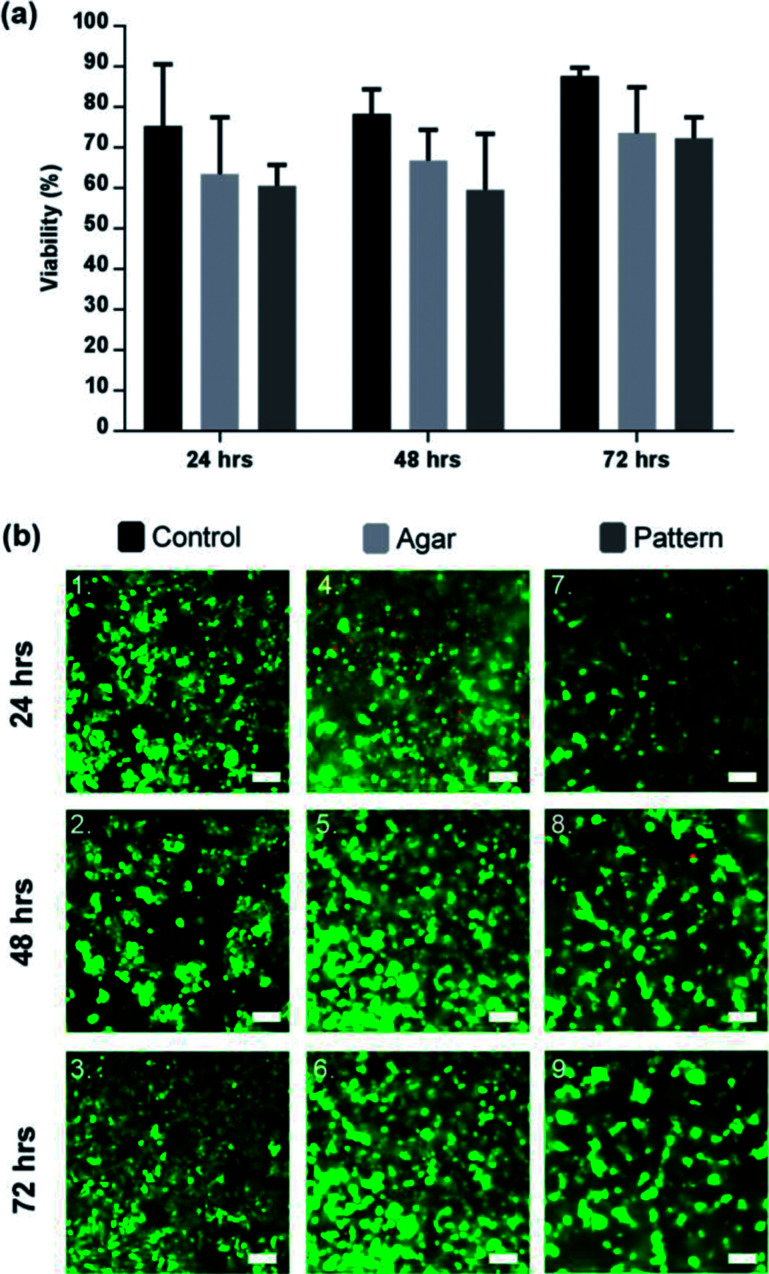
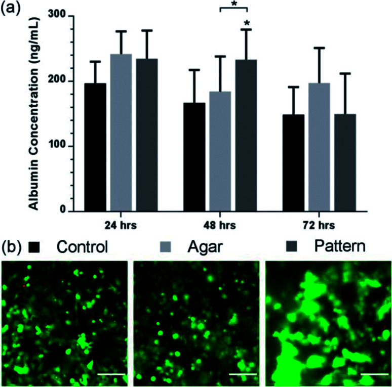
We report upon a novel coplanar dielectrophoresis (DEP) based cell patterning system for generating transferrable hepatic cell constructs, resembling a liver-lobule, in culture. The use of paper reinforced gel substrates provided sufficient strength to enable these constructs to be transfered into 96-well plates for long term functional studies, including in the future, drug development studies. Experimental results showed that hepatic cells formed DEP field-induced structures corresponding to an array of lobule-mimetic patterns. Hepatic viability was observed over a period of 3 days by the use of a fluorescent cell staining technique, whilst the liver specific functionality of albumin secretion showed a significant enhancement due to the layer patterning of cell lines (HepG2/C3A), compared to 2D patterned cells and un-patterned control. This "build and transfer" concept could, in future, also be adapted for the layer-by-layer construction of organs-on-chip in microtitre formats.


Conflict of interest statement

There are no conflicts to declare.

Figures

Fig. 1
Fig. 1. Illustration of the “build and transfer” system. (a) Utilising a single plane biomimetic DEP electrode arrangement, C3A human liver cells where patterned within an agar hydro-gel supported by a 6.2 mm OD, 5 mm ID paper ring. The system was heated during patterning to maintain the liquid state of the agar, then cooled to cure the agar into a gel. Patterning was induced by positive DEP through a 10 MHz, 10 Vpp AC signal. (b) Supported by a paper-ring substrate, the patterned cells were removed by tweezers. (c) Patterned cells were then immediately placed in a 96-well plate for incubation and subsequent biological analysis. Scale bar is 5 mm.
Fig. 2
Fig. 2. The “build and transfer” system setup and operation for patterning liver like structures within an agar gel with a paper substrate. (a) Warmed 1% agar solution with randomly distributed cells at 37 °C was pipetted into volume designated by the gasket. The Peltier warmed the glass substrate on which the electrodes are situated and enclosed with a cover. (b) Hepatic cells were captured by positive DEP forces (F_DEP) induced by a non-uniform electric field (10 MHz, 10 Vpp). (c) Once patterning was complete, the Peltier was switched to cooling mode dropping the temperature of the agar to 18 °C, curing the gel. Once the air temperature reached 25 °C the AC field was switched off. Agar 1% solution enters gel phase at this temperature. (d) The cells patterned by the DEP forces are held in place by the cured agar. The paper substrate containing a biomimetic micro liver is used to manipulate the patterned cells into a culture system.
Fig. 3
Fig. 3. Electrode design. (a) The liver is the largest organ in the body and receives blood from the hepatic portal vein and the hepatic artery. The liver is composed of small units, which can be described in a number of ways. One of the common descriptions is the “classic lobule”. The lobules are approximately hexagonal in cross-section, with the central vein (80 μm diameter) in the centre surrounded radially by sinusoids constructed of hepatocytes. Microelectrode design for inducing DEP forces to pattern cells into a micro liver lobule array. (a) Using a biomimetic electrode design, hepatocytes are patterned into an array 19 lobules in size using DEP forces. The largest gap is 60 μm between the electrodes. The width of the electrodes is 10 μm, thickness 100 nm. (b) Microscope image showing C3A liver cells being held in place by DEP forces to form liver lobule like structures (10 MHz, 10 Vpp for 2 min). Scale bar is 200 μm.
Fig. 4
Fig. 4. Viability of patterned lobule like structures. (a) Viability of 2D control (black), agar control (light grey), and C3A lobule like structures (dark grey) for a period of 72 h. Cell viability in the control was consistently higher than the agar control or pattern by 72 h (87.64 ± 2.0%). Comparatively, the agar control was observed to be 73.6 ± 11.3% and the pattern was 71.3 ± 5.6%. Data represent the mean ± STD for four independent repeats. Statistically there was no significant differences between the 3 sample groups. p > 0.05 (ANOVA). (b) Microscope images of C3A cells after 72 hours of culture. (1–3) 2D sample spread across the surface of the well-plate. (4–6) C3A cells encapsulated within agar gel; cells in close proximity formed small aggregates or spheroids. (7–9) Patterned cells maintained position while forming cords of cells in the shape of the original design. Scale bars 100 μm.
Fig. 5
Fig. 5. Albumin production of lobule like structures. (a) Albumin value (ng mL−1) of control (black), agar control (light grey), and patterned C3A cells (dark grey) over 72 h. Patterned C3A cells had increased albumin secretion after 48 h of culture compared to the control and monolayer control. The control samples show a stable production over the first two days before decreasing. Data represent the mean ± STD for four independent repeats. The (*) indicates statistically significant increase in albumin protein of patterned cells relative to control, and agar 48 h, p ≤ 0.05 (ANOVA). (b) Viability images of control, agar control, and patterned cells. Images are a composite of both live cells (FITC-green) and dead cells (rhodamine – red) images. Scale bars 100 μm.

Similar articles

Cited by

References

    1. Allen J. W. Bhatia S. N. Tissue Eng. 2002;8:725–737. doi: 10.1089/10763270260424097. - DOI - PubMed
    1. Rudolph A. S. Reasor J. Biosens. Bioelectron. 2001;16:429–431. doi: 10.1016/S0956-5663(01)00156-7. - DOI - PubMed
    1. Saltzman W. M. Olbricht W. L. Nat. Rev. Drug Discovery. 2002;1:177–186. doi: 10.1038/nrd744. - DOI - PubMed
    1. Singhvi R. Stephanopoulos G. Wang D. I. Biotechnol. Bioeng. 1994;43:764–771. doi: 10.1002/bit.260430811. - DOI - PubMed
    1. Chiang M.-Y. Hsu Y.-W. Hsieh H.-Y. Chen S.-Y. Fan S.-K. Sci. Adv. 2016;2:e1600964. - PMC - PubMed