. 2018 Mar 12;8(18):9941-9945.
doi: 10.1039/c7ra13237a. eCollection 2018 Mar 5.

β-Carotene: a natural osteogen to fabricate osteoinductive electrospun scaffolds

Affiliations

β-Carotene: a natural osteogen to fabricate osteoinductive electrospun scaffolds

Atiyeh Dabouian et al. RSC Adv. .

Erratum in

Abstract

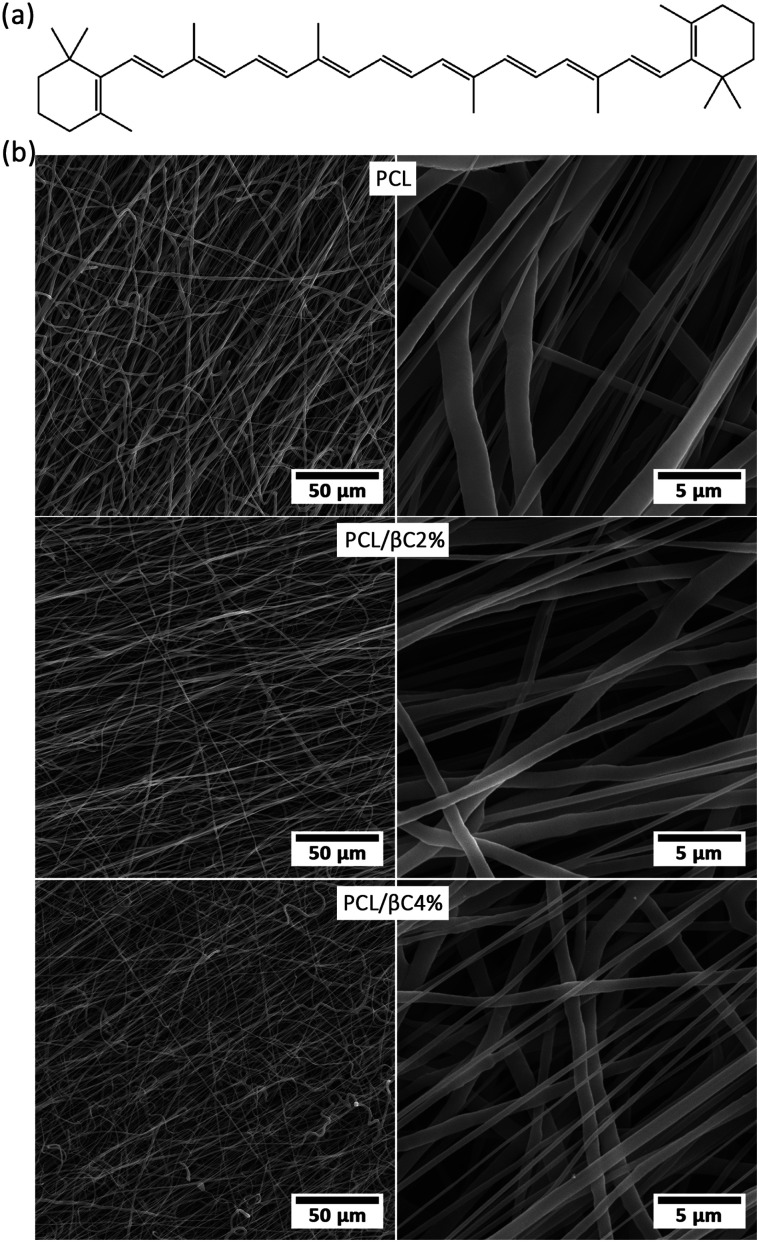
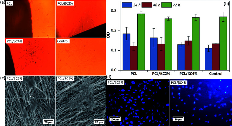
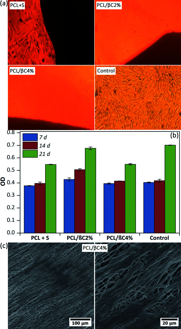
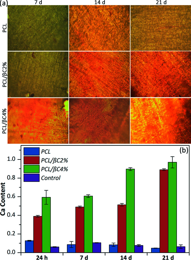
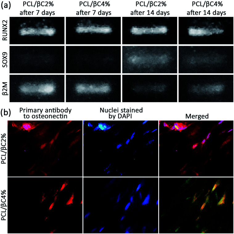
β-Carotene (βC) as a natural osteogenic material was incorporated in PCL electrospun mats to fabricate scaffolds for bone tissue engineering. These scaffolds successfully supported the attachment and proliferation of mesenchymal stem cells (MSCs). Seeded scaffolds were calcinated during 21 days of cell culture in a non-differential medium, which showed the osteodifferentiation of MSCs. Expression of RUNX2, SOX9, and osteonectin proved the osteoinductive effect of incorporated β-carotene on the differentiation of MSCs to osteoblasts without using any external osteogenic differential agent. However, the cells did not pass the early phase of osteogenesis and were still osteochondro-progenitor after 21 days of incubation. Thus, the fabricated fibrous scaffolds are potential candidates for direct bone tissue engineering.


Conflict of interest statement

There are no conflicts of interest to declare.

Figures

Fig. 1
Fig. 1. (a) The molecular structure of β-carotene. (b) SEM images of the fabricated random fibrous mats.
Fig. 2
Fig. 2. (a) Optical microscopic images of MSCs (104 cells per well) on the scaffolds (0.5 × 0.5 cm2) after 24 hours. Tissue culture plate was used as the control. The magnification is 100. (b) The viability of MSCs on the scaffolds up to 76 hours obtained by MTT assay. (c) SEM images of the MSCs attached on the scaffolds after 48 hours. (d) DAPI staining images of the cell nucleus on the scaffolds after 48 hours.
Fig. 3
Fig. 3. (a) Optical microscopic images of differentiated cells (104 cell per well) on the scaffolds (0.5 × 0.5 cm2) after 21 days. Tissue culture plate was used as the control. S was l-ascorbic acid (10 mM) and dexamethasone (1 mM). The magnification is 100. (b) The viability of the cells on the scaffolds up to 21 days obtained by MTT assay. (c) SEM images of the cells on PCL/βC4% scaffold after 14 days.
Fig. 4
Fig. 4. (a) Alizarin red staining of the scaffolds seeded with MSCs cells (104 cells per well). The magnification is 200. (b) Calcium content of the scaffolds seeded with MSCs. Tissue culture plate was used as the control.
Fig. 5
Fig. 5. (a) RT-PCR results for expression of RUNX2 and SOX9 of osteodifferentiated cells (106 cells per well) on scaffolds (0.5 × 0.5 cm2). The expression of β2M was recorded as a control gene. (b) ICC results for expression of osteonectin of osteodifferentiated cells on scaffolds after 21 days. The magnification is 400.

Similar articles

Cited by

References

    1. Johnell O. Kanis J. A. Osteoporosis Int. 2006;17:1726–1733. doi: 10.1007/s00198-006-0172-4. - DOI - PubMed
    1. Wang F. Wang N. Gao Y. Zhou Z. Liu W. Pan C. Yin P. Yu X. Tang M. Life Sci. 2017;174:15–20. doi: 10.1016/j.lfs.2017.03.002. - DOI - PubMed
    1. Liu Y. Wu G. de Groot K. J. R. Soc., Interface. 2010;7:S631–S647. doi: 10.1098/rsif.2010.0115.focus. - DOI - PMC - PubMed
    1. Jun W. Peng W. Dianming J. Hong L. Cong L. Xing L. Xiangyang Q. Yujiang C. Ming L. RSC Adv. 2017;7:54306–54312. doi: 10.1039/C7RA10523A. - DOI
    1. Tao L. Zhonglong L. Ming X. Zezheng Y. Zhiyuan L. Xiaojun Z. Jinwu W. RSC Adv. 2017;7:54100–54110. doi: 10.1039/C7RA06913H. - DOI